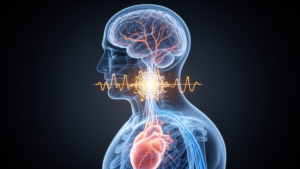
Every heartbeat sends a pulse of blood surging toward the brain, and every shift in body position quietly reshapes how that vital organ is fed. A team of researchers in China has now built an artificial intelligence system that listens to both the heart and the brain at once, using two very different signals to classify the state of cerebral perfusion with remarkable precision. The work, published in Frontiers in Bioengineering and Biotechnology, pairs a wearable near-field coupling sensor that tracks cerebral blood flow with a standard electrocardiogram, then fuses the two streams in a deep learning framework designed specifically for the quirks of each signal. In tests with healthy volunteers undergoing a controlled head-down tilt, the system distinguished between lying flat and being tilted with 95.95 percent accuracy, and it even sorted volunteers by both posture and age group with 79.29 percent accuracy, outperforming several established multimodal models.
The clinical motivation behind the study is stark. Acute ischemic stroke accounts for roughly 70 percent of all strokes and remains a leading cause of death and long-term disability worldwide. The dynamic behavior of cerebral blood flow directly reflects disturbances in cerebral energy metabolism and the extent of neuronal injury, making it a key indicator of stroke progression. Accurate, real-time assessment of blood flow to the brain allows clinicians to track the onset and evolution of ischemia, optimize treatment decisions, and improve prognostic evaluation. Yet the tools currently available for this task are poorly suited to continuous bedside monitoring.
Computed tomography, magnetic resonance imaging, and positron emission tomography provide high spatial resolution and quantitative perfusion information, but their high cost, limited availability, and long acquisition cycles make them impractical for real-time or continuous monitoring. Transcranial Doppler ultrasound offers dynamic measurement but is largely restricted to flow velocity in major cerebral arteries, while near-infrared spectroscopy is constrained by optical attenuation and struggles to reflect deep or whole-brain perfusion. Near-field coupling technology has recently attracted attention as an alternative: it is noninvasive, noncontact, and capable of continuous real-time monitoring. Previous studies have demonstrated its feasibility for detecting intracranial conditions such as cerebral hemorrhage, edema, and ischemic stroke.
The research group behind the new framework had previously developed a wearable NFC-based cerebral blood flow monitoring device capable of simultaneously capturing perfusion changes in both large and small intracranial vessels, validated in healthy volunteers and in patients with cerebrovascular diseases including subarachnoid hemorrhage. Their earlier tilt-based work showed that NFC could characterize dynamic cerebral autoregulation under controlled postural perturbation. But a persistent challenge emerged: under the same tilt paradigm, NFC responses exhibited two distinct trends across individuals, consistent with the substantial inter-individual variability in cerebral perfusion reported across different populations and physiological states. Such variability threatened to undermine the generalizability of any assessment based on cerebral blood flow signals alone.
The solution the team proposed is physiologically elegant. Acute ischemic stroke not only compromises cerebral perfusion but may also involve impaired cerebral autoregulation, and it can precipitate cardiac arrhythmias, reduced heart rate variability, and other forms of cardiac dysfunction. In early ischemic stroke, altered heart rate variability and baroreflex sensitivity have been linked to impaired dynamic cerebral autoregulation and larger infarct volumes. Recent work on brain-heart interactions in stroke emphasizes that cerebrovascular impairment and cardiovascular dysregulation should be considered interdependent processes rather than isolated events. Cardio-cerebral coupling is mediated through interacting hemodynamic, neural, and humoral pathways: postural or pathological changes in venous return, cardiac output, and arterial pressure activate baroreflex-mediated adjustments in heart rate and rhythm, while cerebral autoregulation modifies cerebrovascular tone through myogenic, neurogenic, metabolic, and endothelial mechanisms. Humoral factors including catecholamines, the renin-angiotensin system, and vasopressin further regulate vascular tone and circulating volume over longer time scales.
By incorporating the cardiac regulatory context that the electrocardiogram provides, the researchers aimed to resolve the ambiguity introduced by inter-individual variability in cerebral blood flow responses. The resulting framework consists of two collaborative modules: a Multi-Scale Feature Extractor, or MSFE, and a Depthwise-Separable Cross-Attention Fusion module, or DSCAF. The MSFE employs modality-specific multi-scale convolutional branches with kernel sizes tailored to the temporal characteristics of each signal. The ECG branch uses short kernels of 32 and long kernels of 256 to capture QRS morphologies and RR-interval rhythms, while the NFC-CBF branch uses kernels of 8 and 32 to model impedance peak-valley details and inter-beat fluctuations. Feature channels progressively increase from 128 to 256 to 512 as the network deepens, and Dropout is incorporated to mitigate overfitting. This dual-branch design functions as a physiological filter bank, addressing the limitation of single-scale convolutions that tend to overlook critical temporal structures.
The DSCAF module then integrates depthwise separable convolution with a dual-branch cross-attention mechanism to achieve deep semantic interaction and complementary fusion. Unlike a standard Cross-Transformer that relies on full attention operations over high-dimensional token representations, DSCAF constructs two cross-attention pathways using depthwise-separable projections, a shared bimodal query, and modality-specific key-value representations. This design reduces computational redundancy while preserving bidirectional interactions between the ECG and NFC-CBF streams. Depthwise separable convolution independently extracts local temporal features before integrating them across channels via pointwise convolution, significantly lowering computational complexity for high-dimensional feature maps. Element-wise weighted reconstruction and cross-modal residual connections within each branch produce a unified bimodal representation that explicitly models cross-modal dependencies.
To test the framework, the team built a synchronized monitoring platform combining an NFC-CBF module with an ECG module. The NFC-CBF module used a dual-layer planar spiral electromagnetic coupling sensor, a capacitive digital sensor to convert NFC sensing outputs into a resonant-frequency signal, and a microcontroller that acquired the signal at an effective output rate of 18 Hz before wireless transmission to a host PC. The ECG module recorded signals at a sampling rate of 144 Hz. Data were collected from 20 healthy male volunteers, divided into a young cohort aged 18 to 25 years and a middle-aged cohort aged 35 to 55 years, under two postural conditions: supine and 30-degree head-down tilt. Participants rested for one minute before recording, then simultaneous ECG and NFC-CBF signals were recorded for three minutes supine and four minutes tilted. All participants had no history of cardiovascular, cerebrovascular, or neurological disorders and refrained from caffeine and medication for 24 hours before the experiment.
Preprocessing pipelines suppressed baseline drift and noise in both modalities. For ECG, Butterworth filtering combined with a moving-average kernel removed baseline fluctuations while preserving QRS complex and T wave morphology. For NFC-CBF, wavelet threshold denoising using a hybrid soft- and hard-threshold strategy retained the primary peak and dicrotic wave components with maximal noise suppression. The preprocessed recordings were segmented into non-overlapping 10-second windows, yielding 840 samples for deep learning. Subject-level five-fold cross-validation ensured that all segments from the same participant remained within the same fold, with Z-score parameters calculated from training data only. Two classification tasks were designed: a binary task distinguishing supine from tilted states, and a four-class task jointly distinguishing age group and posture.
The results were striking. In the binary task, the proposed framework achieved an accuracy of 95.95 percent, outperforming ResNet-SE-LSTM at 93.81 percent, the Cross-Transformer Network at 81.07 percent, and Bimodal Feature Fusion at 66.19 percent. The corresponding F1-score and recall were 0.95 and 0.96 respectively, with an area under the ROC curve of 0.973. In the four-class task, the framework reached 79.29 percent accuracy, outperforming the strongest baseline by approximately 3.4 percentage points, with class-wise AUC values ranging from 0.867 to 0.922 and a macro-average of 0.890. Fold-wise paired statistical comparisons confirmed that all improvements over baseline models were statistically significant after Holm correction. Ablation experiments revealed that NFC-CBF alone achieved 92.74 percent accuracy in the binary task while ECG alone reached only 64.76 percent, yet adding the ECG stream improved accuracy over NFC-CBF alone by 3.2 percentage points in the binary task and 11.9 percentage points in the four-class task, supporting the complementary value of cardiac-rhythm information, particularly when posture and age had to be represented simultaneously.
Integrated Gradients attribution analysis complemented these findings at the individual-prediction level. In a representative binary example, NFC-CBF accounted for 68.8 percent of total absolute attribution while ECG accounted for 31.2 percent, consistent with its role as the primary source of cerebral perfusion-related information. In the four-class example, attribution was more evenly distributed at 43.1 percent for ECG and 56.9 percent for NFC-CBF. Simultaneously masking the time points with the highest 10 percent absolute attribution within both modalities reduced the true-class probability by 0.752 and 0.675 in the binary and four-class examples respectively, confirming that temporally localized information from both signals contributed to the predictions. The DSCAF module itself outperformed conventional Add, Multiply, and Concat fusion strategies, achieving improvements of approximately 1.9 and 4.4 percentage points over the best conventional strategy in the binary and four-class tasks respectively, with almost no change in parameter count or model size and an increase of less than 5 percent in training time per epoch.
The physiological interpretation of these results is compelling. Heart rate variability metrics derived from ECG showed a consistent pattern across age groups, with an initial increase after tilting followed by a gradual decline, indicating that tilt-induced cardiac modulation follows a reproducible regulatory trajectory independent of age. In contrast, NFC-CBF-derived parameters such as peak-to-valley distance and systolic slope exhibited substantial inter-individual variability, with some participants showing increases and others decreases after tilt. Younger participants, with greater vascular compliance, showed larger NFC-CBF amplitude variations and stronger phase synchrony between NFC-CBF waveforms and the ECG QRS complex than middle-aged participants. These heterogeneous responses are physiologically consistent with the multifactorial and individually variable regulation of cerebral blood flow during postural perturbation, and they reinforce why relying solely on cerebral blood flow signals may be insufficient for robust assessment across individuals.
The authors are careful to frame the work as proof of concept rather than clinical validation. The sample size was relatively small at 20 participants, all male, and the cohort did not include pathological populations or a broader range of ages. BMI was significantly higher in the middle-aged group, and its potential contribution to the age-group comparison cannot be excluded. Concurrent transcranial Doppler, near-infrared spectroscopy, or arterial blood-pressure measurements were not acquired, so participant-level correspondence between NFC-CBF responses and independent cerebrovascular references could not be evaluated. Acute ischemic stroke represents a more complex and dynamically evolving pathological setting in which cerebral autoregulation, autonomic regulation, vascular compliance, and cardiac function may undergo time-dependent alterations during post-stroke progression, potentially reshaping the temporal coupling between cardiac rhythm and cerebral perfusion. Future studies will need to validate the framework in larger, more diverse, and demographically balanced cohorts including patients at different stages after acute ischemic stroke, incorporate synchronous reference measurements, and pursue hardware-specific evaluation and lightweight optimization before practical deployment on wearable or edge devices.
Even within these constraints, the study establishes a methodological foundation for a new generation of perfusion monitoring. By embedding cardiac rhythm information and cerebral perfusion-related features into a shared learnable representation space, the bimodal framework provides a physiologically grounded functional representation of cardio-cerebral coupling that neither signal could achieve alone. The approach moves beyond handcrafted time-domain, frequency-domain, or time-frequency features that may be insufficient to capture nonlinear inter-modal relationships and complex temporal dependencies in coordinated cardiovascular and cerebrovascular responses. As wearable biosensors continue to advance and deep learning architectures grow more attuned to the heterogeneous temporal characteristics of physiological signals, the prospect of continuous, noninvasive, real-time cerebral perfusion assessment at the bedside moves closer to reality. For a disease that strikes every few seconds somewhere in the world, that prospect carries enormous weight.
Subject of Research: A bimodal deep learning framework for cerebral perfusion assessment using synchronous ECG and near-field coupled CBF signals
Article Title: A bimodal deep learning framework for cerebral perfusion assessment using synchronous ECG and near-field coupled CBF signals
Article References: Wu, S., Wang, J., Wan, C., Li, G., Wang, Z., Li, J., Ning, L., Liu, S., Zhong, J., Luo, J., Yang, S., Jiang, B., Liu, S., Chen, J., Qiao, L., & Chen, Y. (2026). A bimodal deep learning framework for cerebral perfusion assessment using synchronous ECG and near-field coupled CBF signals. Frontiers in Bioengineering and Biotechnology, 14, Article 1912951. https://doi.org/10.3389/fbioe.2026.1912951
Image Credits: AI Generated
DOI: 10.3389/fbioe.2026.1912951
Keywords: bimodal, deep, learning, framework, cerebral, perfusion, assessment, synchronous, near-field, coupled, signals, scientific research
Cite Scienmag News
Cassandra Pierce. (October 17, 2026). Bimodal AI reads heart and brain signals to track cerebral perfusion. Scienmag. https://scienmag.com/bimodal-ai-reads-heart-and-brain-signals-to-track-cerebral-perfusion/
Cassandra Pierce. "Bimodal AI reads heart and brain signals to track cerebral perfusion." Scienmag, 17 October 2026, https://scienmag.com/bimodal-ai-reads-heart-and-brain-signals-to-track-cerebral-perfusion/. Accessed 17 October 2026.
Cassandra Pierce. "Bimodal AI reads heart and brain signals to track cerebral perfusion." Scienmag. October 17, 2026. https://scienmag.com/bimodal-ai-reads-heart-and-brain-signals-to-track-cerebral-perfusion/