


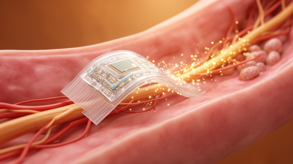
Peripheral nerve injuries leave millions of people with lasting numbness, weakness, and pain, and the gold-standard treatment—autologous nerve grafting—delivers functional recovery that still falls short of normal. Now a comprehensive review published in Biomedical Technology argues that the next leap in nerve repair may come from devices that generate therapeutic electrical stimulation from the body’s own mechanical energy, eliminating the need for bulky external power sources. The review systematically compares three classes of biodegradable nanogenerators—galvanic cells, triboelectric nanogenerators, and piezoelectric nanogenerators—that convert mechanical energy from body motion, respiration, or muscle contraction into electrical output capable of accelerating nerve regeneration.
Electrical stimulation has long been known to enhance peripheral nerve repair, but conventional stimulators require external electrodes and power supplies that complicate clinical use and often demand surgical removal. The review highlights how biodegradable nanogenerators sidestep these limitations by generating stimulation locally, from within the implant itself, and by degrading safely once their therapeutic mission is complete. Galvanic platforms rely on the electrochemical potential between dissimilar metals such as magnesium and molybdenum, producing a steady direct-current electric field of roughly 10 to 100 millivolts per millimeter. Triboelectric devices exploit contact electrification between polymer layers, while piezoelectric platforms generate charge from mechanical deformation of crystalline materials such as polyvinylidene fluoride or polylactic acid.
The review traces the evolution of these devices from early proof-of-concept implants to sophisticated platforms capable of matching autologous nerve grafting in rodent models. A 2020 study reported a fully biodegradable galvanic nerve conduit that produced a steady direct-current field across a 15-millimeter defect in rat sciatic nerve, achieving functional recovery comparable to autologous nerve grafting at 12 weeks. Subsequent work introduced triboelectric platforms that harvest energy from the rhythmic deformation of surrounding tissue, generating alternating-current output that enhanced nerve regeneration across 15-millimeter defects. Piezoelectric conduits have progressed from simple polymer films to multilayered composites incorporating conductive nanofillers, with some platforms achieving functional recovery across 15-millimeter defects at 12 weeks.
The review emphasizes that the therapeutic benefit of these devices depends on far more than raw electrical output. Loaded dose, waveform, frequency, and electrode geometry all shape the biological response, and the review argues that researchers must calibrate these parameters against the specific regenerative context being evaluated. The review also distinguishes the direct electrical stimulation provided by these devices from the indirect mechanical effects of the implant itself, noting that the two can be evaluated separately in controlled experiments. The review further highlights how the biological response varies across the regenerative timeline, with early-phase enhancement of nerve regeneration differing from late-phase effects on functional recovery.
The review identifies galvanic platforms as particularly promising for clinical translation, given their steady direct-current output and their ability to match autologous nerve grafting in rodent models. Triboelectric devices offer the advantage of harvesting energy from the body’s own rhythmic motion, generating alternating-current output that can be tuned to the regenerative context. Piezoelectric conduits have progressed from simple polymer films to sophisticated multilayered composites, with some platforms achieving functional recovery across 15-millimeter defects at 12 weeks. The review argues that the next generation of these devices must integrate wireless activation, calibrated electrical dosing, and safe biodegradation to translate the rodent-model success into clinical practice.
The review concludes that biodegradable nanogenerators represent a promising path toward nerve repair that matches autologous nerve grafting without the limitations of external power sources. The review argues that the next generation of these devices must integrate wireless activation, calibrated electrical dosing, and safe biodegradation to translate the rodent-model success into clinical practice. The review further highlights how the biological response varies across the regenerative timeline, with early-phase enhancement of nerve regeneration differing from late-phase effects on functional recovery. The review concludes that biodegradable nanogenerators represent a promising path toward nerve repair that matches autologous nerve grafting without the limitations of external power sources.
Understanding why electrical cues matter for nerve repair requires looking at what happens inside the injured nerve itself. After a traumatic transection or severe crush, the segment of axon distal to the wound undergoes Wallerian degeneration, a coordinated dismantling process in which the internal cytoskeleton breaks apart and Schwann cells shift from their normal myelinating role into an activated repair state. These repair-phase Schwann cells proliferate and line up inside the residual basal lamina tubes, forming the Bands of Büngner that serve as physical guide rails for regenerating axonal sprouts, while macrophages recruited to the site clear away axonal and myelin debris. Schwann cells also secrete growth factors such as nerve growth factor and glial cell-derived neurotrophic factor, and endothelial cells and fibroblasts help preserve the structural framework of the nerve. Yet even with this endogenous repair machinery in place, axons regrow at only about one to three millimeters per day, and prolonged regeneration allows the repair Schwann cells to lose their activated phenotype, the guidance tubes to collapse, and denervated muscle fibers to atrophy irreversibly.
Exogenous electrical stimulation acts on this repair cascade at several points. At the cellular level, electrical cues influence neuronal membrane polarization and promote calcium influx through voltage-gated calcium channels, setting off downstream signaling through pathways including PI3K/Akt, MAPK/ERK, focal adhesion kinase, and the mechanotransductive regulator YAP. The resulting ion-influx signaling encourages neurite extension, Schwann cell proliferation and migration, secretion of neurotrophic factors, and context-dependent macrophage responses. Electrical stimulation also upregulates regeneration-associated genes such as GAP-43 and enhances BDNF/TrkB signaling through calcium-dependent cAMP signaling. Clinical studies have explored brief perioperative stimulation protocols, such as one hour at 20 hertz, and reported improvements in selected electrophysiological measures of reinnervation, although improved electrophysiology has not always translated into improved functional recovery, a gap that underscores how carefully dosing and outcome measurement must be matched.
The way electrical energy reaches the tissue differs substantially across delivery methods, and this distinction shapes both device design and biological effect. Direct coupling approaches place direct-current or alternating-current electrodes in contact with or implanted within the target nerve, delivering stimulation through physical conduction. Indirect coupling approaches, including capacitive coupling and pulsed electromagnetic fields, generate a homogeneous electromagnetic field between electrodes or convert electromagnetic energy into pulsed stimuli that mimic the body’s own electrophysiological rhythms, inducing electrical effects within cells without direct contact. Each mode carries different implications for electrode geometry, field uniformity, and the degree to which the stimulus can be localized to the repair site rather than dissipating through surrounding tissue.
Conventional electrical stimulation systems, however, depend on externally powered electrodes connected through percutaneous leads or on implantable pulse generators containing rigid, non-degradable batteries. These configurations introduce meaningful clinical risks, including infection and chronic foreign-body inflammation at the hardware site, and they typically require a second invasive surgery to remove the device once therapy is complete. The burden of repeated operations, combined with the challenge of powering implanted electronics over the long regeneration timeline, has motivated the search for self-powered alternatives that draw their energy from the biological environment itself rather than from an external supply.
Among the self-powered strategies, enzymatic biofuel cells have been investigated as a way to harvest energy from physiological chemistry, but their application to neural repair remains constrained by enzyme instability and low power density, limiting their practical utility in this context. By contrast, galvanic, triboelectric, and piezoelectric platforms have each accumulated preclinical evidence in peripheral nerve models, and the review organizes these three approaches within a common analytical framework spanning material selection, device architecture, stimulation characteristics, and biological outcomes. Rather than ranking technologies by unloaded voltage or other isolated device-level metrics, the review emphasizes stimulation dose, experimental conditions, regenerative endpoints, degradation behavior, safety, and translational requirements as the criteria that matter for clinical relevance.
The broader shift in neuroengineering over the past decade reflects a movement away from passive structural bridging toward active, self-powered bioelectronic neuromodulation. This evolution draws on advanced materials science and electrical engineering to develop therapeutic approaches for neurological disorders beyond peripheral nerve repair, including spinal cord, vagus nerve, and sciatic nerve stimulation. The convergence of biodegradable materials with energy-transduction mechanisms represents a departure from earlier generations of nerve guidance conduits made from collagen, polyglycolic acid, or poly(lactic-co-glycolic acid), which provided structural support but could not deliver the dynamic biochemical and bioelectrical cues needed to accelerate axonal elongation, sustain Schwann cell activity, and prevent denervation-induced muscle atrophy.
Establishing the clinical value of self-powered electroactive platforms ultimately depends on demonstrating the delivered stimulation dose with precision, confirming long-term safety as the device degrades, and reproducing functional benefit across independent studies and animal models. The review’s translational framework connects the physics of energy harvesting and device design to the strength of regenerative evidence, aiming to distinguish demonstrated biological effects from device-level performance claims. By framing the comparison around how the energy source and device architecture determine the nature, duration, and controllability of the electrical stimulus, and how those parameters relate to cellular responses and functional outcomes, the review offers a structured basis for evaluating which self-powered platforms are most likely to advance from rodent models toward clinical application in peripheral nerve repair.
Subject of Research: Biodegradable nanogenerators for peripheral nerve repair
Article Title: Self-powered bioelectronics for peripheral nerve regeneration: Electroactive materials, energy transduction, and therapeutic mechanisms
Article References: Kao, F.-C., Pebam, M., Parashar, P., Wang, S.-F., Chiu, P.-Y., Tsai, T.-T., Niu, C.-C., Chen, S.-H., Choi, D., Hsieh, M.-K., & Lin, Z.-H. (2026). Self-powered bioelectronics for peripheral nerve regeneration: Electroactive materials, energy transduction, and therapeutic mechanisms. Materials Today Bio, 41, Article 103734. https://doi.org/10.1016/j.mtbio.2026.103734
Image Credits: AI Generated
DOI: 10.1016/j.mtbio.2026.103734
Keywords: biodegradable nanogenerators, peripheral nerve repair, galvanic cells, triboelectric nanogenerators, piezoelectric nanogenerators, electrical stimulation, nerve regeneration, autologous nerve grafting, Self-powered, bioelectronics, peripheral, nerve
Cite Scienmag News
Denise Maddox. (October 16, 2026). Biodegradable Nanogenerators Turn the Body’s Own Motion Into Nerve Repair. Scienmag. https://scienmag.com/biodegradable-nanogenerators-turn-the-bodys-own-motion-into-nerve-repair/
Denise Maddox. "Biodegradable Nanogenerators Turn the Body’s Own Motion Into Nerve Repair." Scienmag, 16 October 2026, https://scienmag.com/biodegradable-nanogenerators-turn-the-bodys-own-motion-into-nerve-repair/. Accessed 16 October 2026.
Denise Maddox. "Biodegradable Nanogenerators Turn the Body’s Own Motion Into Nerve Repair." Scienmag. October 16, 2026. https://scienmag.com/biodegradable-nanogenerators-turn-the-bodys-own-motion-into-nerve-repair/


