


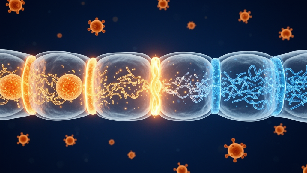
When infection or inflammation strikes the brain, the body’s most formidable defense—the blood-brain barrier—can become its own weak point. A new review published in Frontiers in Malaria synthesizes evidence that two pro-inflammatory cytokines, tumor necrosis factor-alpha (TNF-α) and interferon gamma (IFN-γ), rewire the microRNA landscape of human brain endothelial cells, driving molecular changes that loosen the tight junctions holding the barrier together. The work, led by Nahla Galal Metwally, Sara Mohamad, and Iris Bruchhaus of the Bernhard Nocht Institute for Tropical Medicine in Hamburg, argues that these tiny regulatory RNAs are not bystanders in barrier breakdown but active controllers of it—and that targeting them could open a new therapeutic front in cerebral malaria and other neuroinflammatory diseases.
The blood-brain barrier is built from brain microvascular endothelial cells joined by tight junctions and adherens junctions, protein assemblies that include claudins, occludin, and junctional adhesion molecules anchored to the cytoskeleton by zonula occludens (ZO) scaffolding proteins. This architecture restricts paracellular permeability and keeps the central nervous system insulated from the bloodstream. During infection, activated immune cells release TNF-α and IFN-γ, which act directly on brain endothelial cells. TNF-α increases paracellular permeability in a dose-dependent manner and enhances pathogen penetration, while IFN-γ induces morphological changes, surface expression of class II major histocompatibility complex antigens, and increased permeability to macromolecules. The result is a barrier that admits immune cells, pathogens, and solutes it would normally exclude.
The review details the intracellular signaling cascades through which these cytokines exert their effects. TNF-α binding to its receptors activates the IκB kinase complex, leading to degradation of IκB proteins and nuclear translocation of NF-κB, a transcription factor that switches on genes for adhesion molecules, chemokines, and cytokines. Recent studies show that this inflammatory reprogramming compromises barrier integrity, and that NF-κB inhibition can prevent the morphological remodeling, lipid accumulation, and junctional disruption that follow cytokine stimulation. Mitogen-activated protein kinase pathways—ERK, JNK, and p38—are also activated, contributing to tight junction disruption and matrix metalloproteinase production; TNF-α-induced MMP-9 expression in human cerebral microvascular endothelial cells proceeds through the Ca²⁺/CaMKII/ERK/NF-κB pathway. Glycogen synthase kinase-3β emerges as another key node: its inhibition restores barrier integrity and reduces adhesion molecule expression, partly by modulating miRNA expression and activating the Wnt/β-catenin pathway. Separately, tissue-nonspecific alkaline phosphatase activity, which supports barrier maintenance via the ROCK pathway, is significantly decreased by TNF-α and IFN-γ treatment, promoting cytoskeletal remodeling and barrier loss.
At the heart of the review is the argument that microRNAs—roughly 22-nucleotide non-coding RNAs that bind complementary sequences in the 3′ untranslated regions of target mRNAs, causing translational repression or degradation—translate cytokine signaling into structural barrier failure. Comprehensive profiling shows that TNF-α stimulation of human brain microvascular endothelial cells downregulates 123 miRNAs and upregulates 193. Among the most consistently induced are members of the let-7 family and miR-98, which target the inflammatory chemokines CCL2 and CCL5, suggesting a built-in negative feedback loop to restrain excessive inflammation. In peripheral endothelial cells, TNF-α upregulates miR-31 and miR-17-3p, which target E-selectin and ICAM-1 respectively—findings not yet formally validated in brain endothelium but likely conserved across vascular beds. In brain endothelial cells themselves, miR-125a-5p is upregulated and dampens TNF-α-mediated ICAM-1 expression while reducing monocyte transmigration.
The most extensively characterized cytokine-responsive miRNA is miR-155, which is strongly and rapidly upregulated by combined TNF-α and IFN-γ treatment. Transcriptomic analysis reveals that miR-155 overexpression in human cerebral microvascular endothelial cells downregulates 390 transcripts, with enrichment in processes related to translation, focal adhesion, and inter-endothelial junctional complexes. Its validated targets include annexin-2, claudin-1, dedicator of cytokinesis 1, and syntenin-1. Overexpression of miR-155 mimics cytokine-induced junctional disorganization and permeability increases, while loss of miR-155 in knockout mice reduces barrier leakage in models of neuroinflammation. In contrast, miR-146a—also upregulated by the cytokines and elevated in microvessels of active multiple sclerosis lesions—appears protective, repressing RhoA, NFAT5, IRAK1, and TRAF6 to inhibit NF-κB activation and decrease adhesion molecule expression, thereby reducing T-cell adhesion to the endothelium.
The review emphasizes that timing matters. Early-phase responses within 6 to 12 hours feature rapid upregulation of miR-155 and miR-146a as immediate modulators of inflammatory signaling. Intermediate-phase responses between 12 and 48 hours sustain these miRNAs while additional regulators of junctional proteins emerge, with the balance between barrier-disrupting and barrier-protective species determining the extent of dysfunction. Beyond 48 hours, compensatory upregulation of barrier-protective miRNAs such as let-7 and miR-98 may attempt to restore homeostasis. Dose is equally decisive: lower concentrations of TNF-α and IFN-γ activate caspase-3 and caspase-9, increasing paracellular permeability and redistributing junctional complexes without killing the cells, while higher concentrations fully activate caspase-3/7, triggering endothelial apoptosis and complete barrier breakdown. Combined cytokine treatment also causes redistribution of ZO-1 and VE-cadherin from cell-cell contacts to the cytoplasm, disrupting both tight and adherens junctions.
The authors propose a three-level framework—cytokine-specific, context-specific, and disease-specific—to organize this complexity. Cytokine-specific effects are those directly demonstrated in brain endothelial cells, such as the NF-κB-driven miR-155 and miR-146a responses. Context-specific effects depend on cytokine concentration, exposure duration, and vascular bed: low-dose TNF-α primarily induces reversible tight-junction reorganization in which miR-155 leads, whereas high-dose or prolonged co-exposure shifts toward caspase-mediated breakdown with a partially distinct miRNA signature. Disease-specific effects layer pathogen- or tissue-derived signals on top of the cytokine background. Cerebral malaria adds sequestration-driven endothelial activation and parasite-derived extracellular vesicles; ischemic stroke superimposes hypoxia, inducing miR-210 and miR-212/132; multiple sclerosis combines chronic IFN-γ exposure with autoimmune T-cell signaling. The same miRNA can therefore appear barrier-protective in one disease and barrier-disrupting in another.
Cerebral malaria, caused by Plasmodium falciparum, serves as the review’s translational case study. Comparisons of infected red blood cells and TNF-α on human brain endothelial cells reveal that TNF-α induces strong endothelial activation with only modest permeability increase, whereas infected erythrocytes cause a marked permeability rise and preferentially trigger an endoplasmic reticulum stress response—evidence that parasite-specific signaling is a major driver of barrier disruption beyond cytokines alone. Infected erythrocyte-derived extracellular vesicles deliver miR-451a and other Ago2-miRNA complexes into endothelial cells, reducing caveolin-1 expression and altering barrier function, and are also enriched in let-7i, miR-150, and miR-146a—the same miRNAs that host cytokines induce endogenously. In experimental cerebral malaria in mice, circulating exosomal miR-155 rises over the disease course, miR-155 deficiency preserves barrier integrity, and antagomir-155 reduces vascular leak in microvessels exposed to sera from Ugandan children with cerebral malaria. Yet in human cohorts, circulating miR-155 has not consistently distinguished cerebral malaria from uncomplicated disease, and miR-451a’s abundance in erythrocytes generally complicates its specificity as a biomarker—discrepancies the authors attribute to differences between murine CD8⁺ T-cell-driven pathology and human sequestration-dominated disease, cohort age and genotype variation, and the compartment in which miRNAs are measured.
The therapeutic implications are substantial. The review outlines three strategies: inhibiting barrier-disrupting miRNAs such as miR-155 with antisense oligonucleotides or locked nucleic acid inhibitors; delivering barrier-protective miRNAs such as miR-98, let-7, or miR-146a using synthetic mimics or exosome-based systems, building on proof of concept from bone marrow stromal cell-derived exosomal miR-21a-5p, which upregulates claudin-5, occludin, and ZO-1 in stroke models; and modulating upstream signaling pathways such as NF-κB or GSK3β to shift the miRNA balance toward protection. Major challenges remain, including delivering miRNA therapeutics across the very barrier they aim to protect, minimizing off-target effects from miRNAs that can regulate hundreds of genes, and determining optimal timing—early intervention before severe barrier breakdown is likely most effective. The authors call for single-cell sequencing to map miRNA heterogeneity across endothelial populations, mechanistic validation of additional targets, investigation of interactions with long non-coding and circular RNAs, and comparative studies of species differences. If those hurdles can be cleared, the microRNA networks that cytokines exploit to dismantle the blood-brain barrier may become the very levers clinicians pull to keep it intact.
Subject of Research: Cytokine-induced microRNA changes in brain endothelial cells and their role in blood-brain barrier dysfunction
Article Title: How do pro-inflammatory cytokines alter miRNA profiles in brain endothelial cells? Mechanisms, consequences, and therapeutic strategies for blood-brain barrier protection
Article References: How do pro-inflammatory cytokines alter miRNA profiles in brain endothelial cells? Mechanisms, consequences, and therapeutic strategies for blood-brain barrier protection. (n.d.). https://doi.org/10.3389/fmala.2026.1849845
Image Credits: AI Generated
DOI: 10.3389/fmala.2026.1849845
Keywords: blood-brain barrier, microRNAs, TNF-alpha, IFN-gamma, cerebral malaria, neuroinflammation, brain endothelial cells, miR-155, pro-inflammatory, cytokines, alter, miRNA
Cite Scienmag News
Cassandra Pierce. (October 16, 2026). Cytokine-Driven microRNA Shifts in Brain Endothelial Cells Offer New Route to Protect the Blood-Brain Barrier. Scienmag. https://scienmag.com/cytokine-driven-microrna-shifts-in-brain-endothelial-cells-offer-new-route-to-protect-the-blood-brain-barrier/
Cassandra Pierce. "Cytokine-Driven microRNA Shifts in Brain Endothelial Cells Offer New Route to Protect the Blood-Brain Barrier." Scienmag, 16 October 2026, https://scienmag.com/cytokine-driven-microrna-shifts-in-brain-endothelial-cells-offer-new-route-to-protect-the-blood-brain-barrier/. Accessed 16 October 2026.
Cassandra Pierce. "Cytokine-Driven microRNA Shifts in Brain Endothelial Cells Offer New Route to Protect the Blood-Brain Barrier." Scienmag. October 16, 2026. https://scienmag.com/cytokine-driven-microrna-shifts-in-brain-endothelial-cells-offer-new-route-to-protect-the-blood-brain-barrier/


