


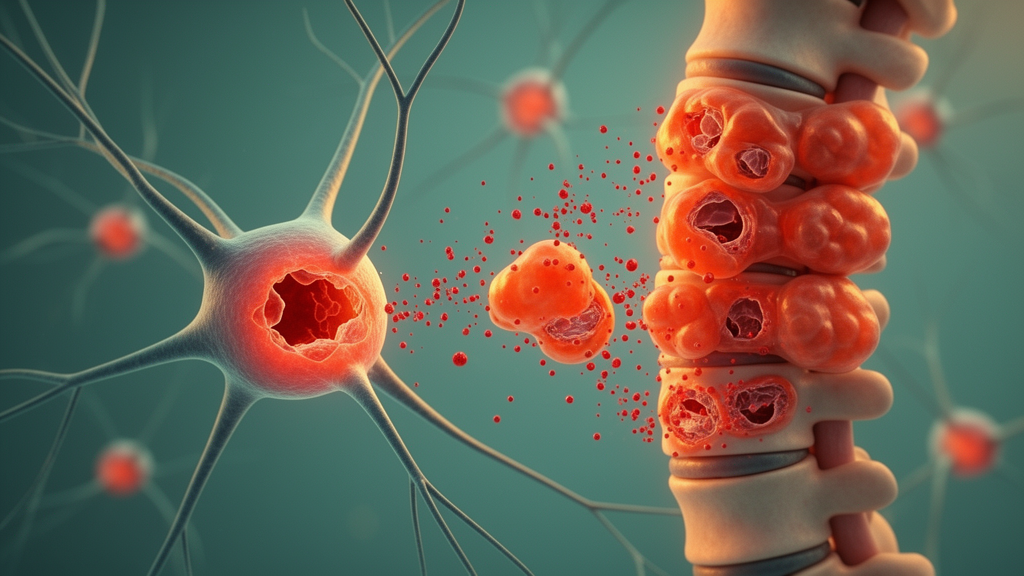
When a stroke cuts off blood flow to the brain, when a hemorrhage floods neural tissue with blood, or when a spinal cord is crushed in an accident, the initial damage is only the beginning of the story. In the hours and days that follow, injured cells of the central nervous system unleash a cascade of secondary processes, including metabolic collapse, oxidative stress, rampant inflammation, and a set of genetically programmed cell death routines that can wipe out neighboring tissue that survived the original insult. A new review published in Frontiers in Molecular Neuroscience brings together two of the most consequential threads in this cascade, mitochondrial dysfunction and a hybrid form of inflammatory cell death known as PANoptosis, and argues that the power plants of the cell may be the upstream switch that triggers the entire lethal program.
The review, which surveys evidence from ischemic stroke, intracerebral hemorrhage, traumatic brain injury, and spinal cord injury, positions mitochondria near the center of acute central nervous system injury for a straightforward reason: these organelles perform multiple jobs at once, and when they fail, they fail catastrophically. Mitochondria generate the ATP that neurons and glial cells need to maintain ion gradients and survive, but they also produce reactive oxygen species as a byproduct of respiration, regulate their own dynamics through constant fission and fusion, and are selectively degraded through mitophagy when damaged. In acute injury, impaired bioenergetics starves cells, excessive mitochondrial reactive oxygen species oxidize proteins, lipids, and DNA, abnormal fission fragments the network, defective mitophagy allows damaged organelles to accumulate, and mitochondrial DNA escapes into the cytoplasm, where it is mistaken for an invading pathogen.
That last point is where the connection to PANoptosis becomes concrete. Mitochondrial DNA, cardiolipin, and other mitochondrial molecules act as damage-associated molecular patterns, or DAMPs, that are normally sequestered inside the organelle. When mitochondrial membranes rupture, these molecules spill into the cytosol and activate DNA-sensing pathways and inflammasome complexes, the molecular assemblies that ignite inflammatory cell death. The review describes how this mitochondrial stress creates conditions that favor PANoptotic convergence: rising mitochondrial reactive oxygen species and released DAMPs activate inflammasome signaling, while sustained activity of receptor-interacting protein kinases and caspases links the separate death pathways together into a single coordinated execution.
PANoptosis itself is a relatively recent concept in cell biology. Classical descriptions of cell death treated pyroptosis, apoptosis, and necroptosis as distinct programs, each with its own molecular machinery. Pyroptosis is a highly inflammatory form of death driven by gasdermin pores that rupture the cell membrane, apoptosis is a quieter, caspase-mediated dismantling of the cell, and necroptosis is a lytic death executed by the kinase pair RIPK1 and RIPK3 together with the pseudokinase MLKL. PANoptosis collapses these boundaries. In this model, a multiprotein complex called the PANoptosome assembles in response to specific danger signals and simultaneously engages components of all three pathways, so a single cell can die with features of pyroptosis, apoptosis, and necroptosis at once. The result is a form of death that is both genetically regulated and violently inflammatory, a combination particularly destructive in the confined space of the skull or spinal canal.
The review is careful to distinguish between direct evidence of PANoptosis and studies that merely show coordinated changes in individual death pathways, and this distinction matters for interpreting the literature on acute brain and spinal cord injury. Many experiments in stroke, hemorrhage, and trauma models report simultaneous activation of inflammasome components, caspases, and necroptotic kinases, which is consistent with PANoptotic signaling but does not by itself prove that a defined PANoptosome assembled in the injured tissue. The authors conclude that complete causal evidence linking mitochondrial damage to defined PANoptosome assembly remains limited in acute central nervous system injury, a candid assessment that tempers the enthusiasm surrounding the concept while still supporting its biological plausibility.
Within the four injury types the review covers, the mitochondrial picture varies in detail but shares a common architecture. In ischemic stroke, interrupted blood flow deprives mitochondria of oxygen and substrates, and reperfusion then floods the tissue with a burst of reactive oxygen species that damages the electron transport chain and opens the mitochondrial permeability transition pore. In intracerebral hemorrhage, blood products such as hemoglobin and iron impose oxidative stress directly on mitochondrial membranes. In traumatic brain injury and spinal cord injury, mechanical forces disrupt axons and trigger calcium overload, excitotoxicity, and sustained mitochondrial permeabilization that propagates secondary damage across networks of connected neurons. In all four conditions, the review finds evidence of disturbed fission and fusion dynamics, impaired mitophagy, and release of mitochondrial DAMPs that feed inflammatory signaling.
Two newer areas of mitochondrial biology also feature in the analysis. Mitochondria-derived vesicles are small carriers that bud off from mitochondria and can transport damaged components away for degradation or deliver mitochondrial material to other compartments, and their behavior in acute injury may influence both quality control and inflammatory signaling. Mitochondrial transfer, the movement of intact mitochondria between cells, has attracted attention as a potential rescue mechanism, with experimental studies exploring whether healthy mitochondria delivered to injured neural tissue can restore bioenergetics. The review evaluates these processes alongside classical targets such as oxidative stress, dynamics, and mitophagy, and assesses therapeutic strategies aimed at each of them.
On the therapeutic side, the review surveys strategies that target mitochondrial oxidative stress with antioxidants and reactive oxygen species scavengers, agents that modulate mitochondrial fission and fusion, approaches that enhance mitophagy to clear damaged organelles, interventions that stimulate mitochondrial biogenesis to replace dysfunctional networks, and blockers of mitochondrial DAMP signaling that would prevent released mitochondrial molecules from activating inflammasomes. Mitochondrial transplantation and transfer represent the most direct intervention, attempting to supply functional organelles to energy-starved cells. The unifying logic is that mitochondria sit upstream of the death and inflammation cascades, so repairing or protecting them could prevent the downstream activation of PANoptotic signaling rather than trying to inhibit each death pathway individually after it has already been triggered.
The upstream positioning of mitochondria is precisely what makes them attractive as an intervention point, and the review argues that current findings support this promise. A single upstream target that influences bioenergetics, oxidative stress, inflammatory priming, and cell death execution could address several secondary injury mechanisms at once, which is a significant advantage in a field where most neuroprotective agents tested in clinical trials have failed to improve outcomes. At the same time, the authors emphasize that clarifying cell-specific and time-dependent mechanisms will be essential for translating the framework into effective neuroprotective strategies. Neurons, astrocytes, microglia, and infiltrating immune cells respond differently to mitochondrial stress, and the window during which mitochondrial interventions can alter the course of injury may be narrow and stage-specific.
For researchers in neurotrauma and stroke, the review offers a map of where the evidence stands and where the gaps lie. The convergence of mitochondrial dysfunction and PANoptosis-related signaling is well supported as a correlation across injury models, but the field still needs direct demonstrations of PANoptosome assembly in injured central nervous system tissue, causal tests that mitochondrial damage is necessary and sufficient for PANoptotic death, and cell-type-resolved analyses that identify which populations execute which death programs at which time points. Filling those gaps, the authors suggest, will determine whether mitochondria can move from a promising upstream correlate to a validated therapeutic target in the acute injuries that cause so much disability worldwide.
Subject of Research: Mitochondrial dysfunction and PANoptosis-related signaling in acute central nervous system injuries
Article Title: Mitochondrial dysfunction and PANoptosis-related signaling in acute central nervous system injuries: mechanistic convergence and therapeutic opportunities
Article References: Zhang, H., Tu, R., Zhang, D., Tian, H., & Yin, H. (2026). Mitochondrial dysfunction and PANoptosis-related signaling in acute central nervous system injuries: mechanistic convergence and therapeutic opportunities. Frontiers in Molecular Neuroscience, 19, Article 1982074. https://doi.org/10.3389/fnmol.2026.1982074
Image Credits: AI Generated
DOI: 10.3389/fnmol.2026.1982074
Keywords: mitochondria, PANoptosis, ischemic stroke, intracerebral hemorrhage, traumatic brain injury, spinal cord injury, mitochondrial DNA, inflammasome, mitophagy, reactive oxygen species, mitochondrial transfer, neuroprotection
Cite Scienmag News
Cassandra Pierce. (October 16, 2026). When Mitochondria Fail, Inflammatory Cell Death Follows in Brain and Spinal Cord Injury. Scienmag. https://scienmag.com/when-mitochondria-fail-inflammatory-cell-death-follows-in-brain-and-spinal-cord-injury/
Cassandra Pierce. "When Mitochondria Fail, Inflammatory Cell Death Follows in Brain and Spinal Cord Injury." Scienmag, 16 October 2026, https://scienmag.com/when-mitochondria-fail-inflammatory-cell-death-follows-in-brain-and-spinal-cord-injury/. Accessed 16 October 2026.
Cassandra Pierce. "When Mitochondria Fail, Inflammatory Cell Death Follows in Brain and Spinal Cord Injury." Scienmag. October 16, 2026. https://scienmag.com/when-mitochondria-fail-inflammatory-cell-death-follows-in-brain-and-spinal-cord-injury/


