


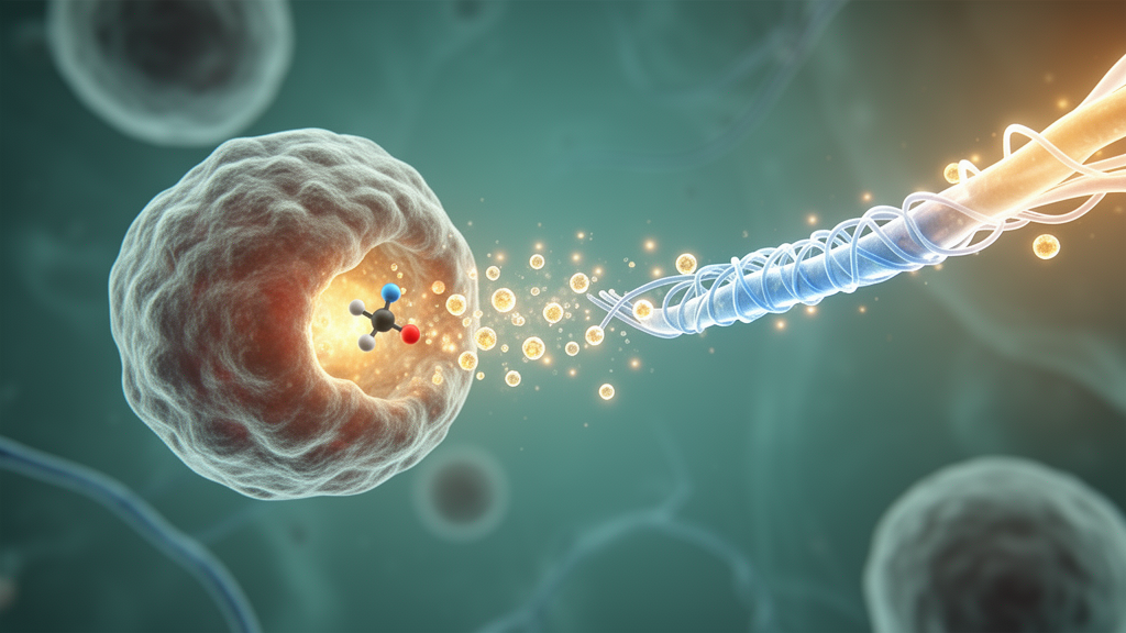
Myelin, the fatty insulation wrapped around nerve fibers by oligodendrocytes, is among the most vulnerable casualties of diseases like multiple sclerosis. When the sheath is stripped away, axons are exposed to a barrage of stressors and slowly degenerate, and the brain’s own repair crews often fail to complete the job. Now researchers in Italy report a mechanism that could explain why one class of immunomodulatory drugs, Bruton’s tyrosine kinase inhibitors, appears to do more than quiet inflammation: it may actively coax damaged neural circuits to rewrap themselves, with tiny membrane bubbles shed by microglia acting as the couriers of repair.
In a study published in Frontiers in Cellular Neuroscience, a team led by Matteo Tartaglia and Myriam Catalano of Sapienza University of Rome, working with colleagues at the CNR Institute of Biomedical Technologies and IRCCS Neuromed, built a laboratory system that reproduces demyelination and recovery in a dish. The work centers on ibrutinib, an irreversible Bruton’s tyrosine kinase inhibitor best known as a blood cancer drug, and asks a deceptively simple question: when ibrutinib helps myelin repair, is it acting on the neurons, on the immune cells, or on the messages passed between them?
To answer it, the researchers needed a model that could sustain real myelination. They isolated spinal cord and brain cortex tissue from postnatal mouse pups aged zero to two days, dissociating the tissue into primary mixed cultures that contained neurons, oligodendrocyte lineage cells, microglia, and astrocytes in proportions resembling the native tissue. By day 35 in vitro, the cultures were floridly myelinated: roughly 59 percent of spinal cord neurons and 52 percent of cortical neurons carried myelin basic protein around their neurites, verified in three dimensions using z-stack confocal imaging and a strict colocalization criterion. Importantly, the team found that the window for study was narrow. By day 45, myelination declined substantially in both culture types, so day 35 became the sweet spot for perturbation experiments.
The demyelinating insult came from lysophosphatidylcholine, or LPC, a detergent-like lipid that disrupts the myelin sheath directly, triggering a secondary immune response that mimics key pathological features of multiple sclerosis. Twenty-four hours of LPC treatment slashed myelination in spinal cord cultures from about 62 percent to 29 percent, and in cortical cultures from roughly 53 percent to 23 percent. Over the following week, the cultures tried to heal themselves. The cortical cultures managed an essentially complete recovery on their own, but the spinal cord cultures only partially rebounded, reaching about 50 percent of neurons remyelinated. That incomplete spontaneous repair made the spinal cord preparations the ideal test bed for a drug that might accelerate the process.
Ibrutinib delivered that acceleration. Applied one day after LPC exposure for 24 hours, the drug drove spinal cord cultures to remyelination levels indistinguishable from healthy controls, while offering no additional benefit in the cortex, where spontaneous repair was already sufficient. This pattern matters mechanistically: Bruton’s tyrosine kinase is a cytoplasmic signaling enzyme expressed in immune cells and, within the central nervous system, mainly by microglia and other CD11b-positive myeloid cells, but not by oligodendrocytes themselves. Any pro-remyelinating effect of blocking it therefore cannot be a direct action on the myelin-forming cells. It must be mediated through the immune compartment, most plausibly through microglia, which adopt an inflamed phenotype after demyelination and can either amplify damage or support repair.
To pin down that mediation, the team turned to BV2 cells, a murine microglia-like cell line. Since LPC alone did not induce a detectable inflammatory program in these cells, which lack neurons and oligodendrocytes, the researchers used lipopolysaccharide, or LPS, to impose a strongly pro-inflammatory state. They then collected the small extracellular vesicles, membrane-enclosed nanoparticles typically around 160 nanometers in diameter, that the cells released into the culture medium. Extracellular vesicles have become a focus of neuroimmunology because their cargo reflects the state of the donor cell and can reprogram recipient cells. Earlier work has shown that microglial vesicles can influence oligodendrocyte precursor cells and astrocytes at myelin lesions, sometimes helping and sometimes hindering repair.
The results were striking. Ibrutinib reduced the sheer number of vesicles shed by inflamed BV2 cells without changing their size profile, suggesting it interferes with the vesicle-release machinery itself. More importantly, it changed what the vesicles carried. Quantitative PCR revealed that vesicles from inflamed microglia treated with ibrutinib contained significantly more miR-223, a microRNA with established regenerative credentials, while levels of the pro-inflammatory miR-155 were unchanged. miR-223 has been shown in previous studies to promote debris clearance and regenerative myeloid phenotypes in the demyelinated nervous system, and it is upregulated in patients with multiple sclerosis compared with healthy individuals. When the researchers applied vesicles from LPS-plus-ibrutinib-treated microglia to demyelinated spinal cord cultures, remyelination improved just as it had with direct ibrutinib treatment. Crucially, the benefit persisted even when endogenous vesicle release in the cultures was blocked with calpeptin, confirming that the effect came from the applied vesicles rather than from modulating the recipient cells’ own vesicle traffic.
The final piece of the puzzle pointed to a specific molecular target. Searching the miRCarta database, the team identified stathmin-1, encoded by the STMN1 gene, as a predicted target of miR-223. Stathmin-1 is a microtubule-destabilizing protein that inhibits tubulin polymerization and stiffens the cytoskeleton, and it is overexpressed in oligodendrocytes from patients with multiple sclerosis, where it may obstruct the cellular remodeling that remyelination demands. In spinal cord cultures recovering from LPC injury, ibrutinib treatment significantly reduced STMN1 expression. The emerging model is elegant: blocking Bruton’s tyrosine kinase in inflamed microglia reshapes the cargo of their extracellular vesicles, enriching them with miR-223, which then reaches oligodendrocyte lineage cells and suppresses a cytoskeletal brake on repair.
The study also carries a practical innovation. By adapting an existing protocol to postnatal mice rather than rat embryos, the researchers created a myelinating culture system that better aligns with the 3Rs principles for animal research while faithfully reproducing the cellular composition of spinal cord and cortex. The nearly three-to-one ratio of non-neuronal to neuronal cells in their cultures matched estimates from mouse, monkey, and human nervous system tissue, lending the model physiological credibility. Whether the postnatal system reveals dynamics that embryonic cultures mask, such as the age-dependent decline in myelination after day 40, remains to be explored in disease models.
For the multiple sclerosis field, the implications are timely. Several Bruton’s tyrosine kinase inhibitors are in clinical development, with phase 2 trials of brain-penetrant candidates already reporting safety and efficacy signals in relapsing disease. Those drugs are thought to work largely by damping B cell and microglial activation, but this study suggests a second, reparative arm: beyond suppressing inflammation, BTK inhibition may actively retune microglial communication to favor remyelination. The authors are careful to frame their findings as preliminary, derived from in vitro systems and a microglia-like cell line. Yet the demonstration that the pro-repair signal travels in vesicles, and that a specific microRNA-protein axis may mediate it, opens a concrete path toward therapies that do not merely halt demyelination but hand the nervous system the molecular tools to rebuild what it has lost.
Subject of Research: Ibrutinib-mediated acceleration of remyelination through microglial extracellular vesicle signaling
Article Title: Neuron myelination is accelerated by Bruton’s tyrosine kinase inhibition via microglial extracellular vesicles
Article References: Tartaglia, M., Ioni, A., De Luca, M., Vilardo, L., D’Agnano, I., Conte, A., Limatola, C., & Catalano, M. (2026). Neuron myelination is accelerated by Bruton’s tyrosine kinase inhibition via microglial extracellular vesicles. Frontiers in Cellular Neuroscience, 20, Article 1864654. https://doi.org/10.3389/fncel.2026.1864654
Image Credits: AI Generated
DOI: 10.3389/fncel.2026.1864654
Keywords: multiple sclerosis, remyelination, ibrutinib, Bruton's tyrosine kinase, microglia, extracellular vesicles, miR-223, stathmin-1, oligodendrocytes, demyelination, neuroinflammation, spinal cord cultures
Cite Scienmag News
Cassandra Pierce. (October 16, 2026). BTK Inhibitor Ibrutinib Speeds Remyelination via Reprogrammed Microglial Vesicles. Scienmag. https://scienmag.com/btk-inhibitor-ibrutinib-speeds-remyelination-via-reprogrammed-microglial-vesicles/
Cassandra Pierce. "BTK Inhibitor Ibrutinib Speeds Remyelination via Reprogrammed Microglial Vesicles." Scienmag, 16 October 2026, https://scienmag.com/btk-inhibitor-ibrutinib-speeds-remyelination-via-reprogrammed-microglial-vesicles/. Accessed 16 October 2026.
Cassandra Pierce. "BTK Inhibitor Ibrutinib Speeds Remyelination via Reprogrammed Microglial Vesicles." Scienmag. October 16, 2026. https://scienmag.com/btk-inhibitor-ibrutinib-speeds-remyelination-via-reprogrammed-microglial-vesicles/


