


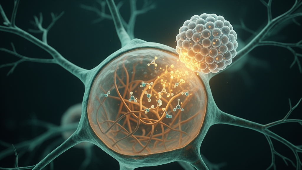
Neuroscientists at Northwestern University have refined a decades-old lipid technique into a practical tool for smuggling fully functional antibodies into living nerve cells, and in doing so they have stumbled onto an unexpected biological finding: a form of tau protein long branded as an Alzheimer’s disease marker appears to be actively involved in keeping neuronal processes intact. The study, published in Frontiers in Dementia, describes how cationic liposomal delivery, or CLD, can flood entire populations of cultured neurons with intact antibodies within an hour, without detectable toxicity, and how one such antibody—PHF-1, which recognizes tau phosphorylated at the serine 396 and 404 residues—triggered rapid neurite retraction and cell-body rounding when released inside the cells.
The problem the researchers set out to solve is one of the most stubborn technical bottlenecks in cell biology. Antibodies are superb molecular recognition machines, capable of distinguishing protein conformations, aggregation states, and individual post-translational modifications with extraordinary precision. But the plasma membrane is an impenetrable barrier to these Y-shaped proteins. Traditional workarounds each carry significant drawbacks. Microinjection and electroporation can introduce antibodies into cells but are slow, laborious, and damaging, making them poorly suited for treating large populations. Genetically encoded intrabodies can be expressed inside cells, but they demand viral vectors or transfection, require days to weeks of expression, and typically perturb all forms of a target protein at once rather than a single modified species. In many cases, antibodies that do get taken up remain trapped in endosomes, never reaching their cytoplasmic targets.
The Northwestern team, led by Kirsten Viola, Brett Chromy, Mary Lambert, and William Klein of the Department of Neurobiology, turned to a cationic lipid with an unusual pedigree. The molecule, 1,2-dioleoyl-sn-glycero-3-ethylphosphocholine, abbreviated DOPC+, was originally developed as a gentle vehicle for DNA transfection. The CLD procedure is remarkably simple and takes less than an hour from start to finish. First, the lipid is dried into a thin film inside a glass tube under a stream of nitrogen or argon. Next, a small volume of antibody solution is added to resuspend the film, and the mixture is vortexed into a milky suspension of multi-lamellar vesicles. A brief sonication step breaks these large vesicles down into small unilamellar liposomes roughly 50 nanometers in diameter, as confirmed by atomic force microscopy. Finally, the antibody-lipid complexes are diluted into serum-free culture medium and applied to the cells, where delivery occurs in under thirty minutes.
To validate the platform, the researchers first tested whether CLD could ferry small molecules across the membrane. Fluorescent BODIPY-phallacidin, a probe that binds actin filaments, was delivered into cultured 3T3 fibroblasts and B103 rat neuroblastoma cells, where it lit up stress fibers and actin bundles at the tips of filopodia within twenty minutes. A synthetic peptide derived from the N-terminal domain of the tyrosine kinase Fyn was likewise delivered into B103 cells and dispersed homogeneously through the cytoplasm. Dextrans ranging from 10 to 70 kilodaltons also crossed successfully. The critical control came from mixing unencapsulated antibody with empty liposomes: essentially no intracellular labeling resulted. The antibody must be incorporated into the lipid complex itself; simply bathing cells in antibody alongside empty vesicles accomplishes nothing.
The decisive experiments involved full-length antibodies. When N1E-115 mouse neuroblastoma cells were incubated with liposomes loaded with Fyn3 antibody, the resulting staining pattern across the population was nearly identical to that obtained by conventional immunocytochemistry of fixed, permeabilized cells. Antibodies against vinculin and paxillin—two components of focal adhesions, the protein complexes that anchor cells to their substrate—were delivered into B103 cells and correctly localized to discrete puncta at the tips of growth cones, exactly where these proteins are known to reside. Because focal contacts have such a characteristic, spatially restricted distribution, they served as a rigorous test of whether delivered antibodies retain their targeting function in the living cytoplasm. They did, without exception.
With the delivery system validated, the team deployed their Alzheimer’s-relevant cargo. PHF-1 is a monoclonal antibody raised against paired helical filament tau, the hyperphosphorylated species that accumulates in the neurofibrillary tangles that define Alzheimer’s disease progression. It binds tau phosphorylated at serines 396 and 404, epitopes widely used as pathological biomarkers in tissue staining and in emerging fluid diagnostics. Yet the same epitope is not exclusively pathological. Earlier work had shown that PHF-1-reactive tau appears transiently in the developing nervous system—both in human fetal brain and in embryonic chick retina—where tau phosphorylation is dynamically regulated during axon formation. The B103 and N1E-115 cell lines used in the study constitutively express the PHF-1 epitope, making them convenient models.
When encapsulated PHF-1 was delivered into B103 cells, it produced filamentous staining patterns identical to those seen with traditional fixation-and-permeabilization labeling, confirming that the antibody had escaped the endosomal route and accessed its cytoskeletal target. Delivery into primary chick retinal neurons—an important demonstration, since these post-mitotic cells cannot be expanded and are far more fragile than immortalized lines—was similarly efficient, with virtually every neuron in the culture taking up the antibody. Crucially, cells tolerated the procedure well: cultures incubated with the complexes for more than twenty-four hours showed no detectable toxicity, and up to 100 micrograms of DOPC+ per milliliter of medium was tolerated by rat hippocampal neurons, chick retinal neurons, and the B103 line alike.
The surprise came when the researchers monitored living B103 cultures by time-lapse imaging after PHF-1 delivery. Within approximately three hours, cells receiving active PHF-1 exhibited dramatic neurite retraction and cell-body rounding—their slender processes withdrew, and the neurons collapsed into rounded shapes. Cultures receiving heat-inactivated PHF-1, empty vehicle liposomes, or other intracellular antibodies showed no such response. This specificity rules out a nonspecific consequence of liposomal delivery or of antibody accumulation inside cells. Instead, it points to a direct functional consequence of antibody binding: engaging the PHF-1-reactive tau pool apparently disrupts processes that maintain neuronal architecture. The authors are careful about interpretation. They note that the mechanism remains unknown—binding might interfere with physiological interactions between phosphorylated tau and the cytoskeleton, alter tau’s conformation, trafficking, or subcellular organization, or, because antibodies are multivalent, crosslink tau species in ways that perturb their function. Nor do the data imply that phosphorylation at Ser396/404 is itself uniquely required for neurite maintenance; the phenotype implicates the PHF-1-reactive population as a whole.
What the finding does suggest is provocative: a tau phospho-form celebrated as a hallmark of neurodegeneration may also perform a physiological job in healthy or developing neurons, one that becomes dysregulated in disease. This dual identity fits a broader theme in modern proteomics. Proteoforms—distinct versions of a protein generated by post-translational modifications such as phosphorylation, acetylation, truncation, or ubiquitination—can carry opposite biological activities, and recent mass-spectrometry efforts have mapped increasingly detailed tau proteoform landscapes in human brain. But defining what each proteoform actually does in a living cell has lagged far behind cataloguing what it is. Genetic approaches perturb the entire protein, not a single modified species.
CLD offers a way to close that gap. Because it delivers pre-formed, full-length antibodies acutely and broadly across a culture, it converts antibodies from passive imaging reagents into active perturbation tools, allowing researchers to interrogate specific endogenous proteoforms in their native cellular context within hours rather than weeks. In the present study, PHF-1 was initially little more than model cargo; its introduction revealed a robust and previously unappreciated phenotype. The same strategy could now be applied to disease-associated species across a range of neurodegenerative disorders. The authors caution that key questions remain—whether the neurite retraction is reversible, what downstream cytoskeletal changes mediate it, and whether the same response occurs in intact neural circuits in vivo. But the demonstration that a simple cationic lipid can deliver functional antibodies safely into populations of living neurons, and that doing so can expose an unexpected function of an Alzheimer’s-linked tau species, marks CLD as a versatile new window onto the hidden workings of the neuronal interior.
Subject of Research: Cationic liposomal delivery of functional antibodies into living neurons and the role of phosphorylated tau in neurite integrity
Article Title: Safe and efficient intracellular delivery of functional antibodies into cultured neural cells shows a role for Alzheimer’s-relevant tau phosphorylation in cell integrity
Article References: Viola, K. L., Chromy, B. A., Nowar, R., Lambert, M. P., & Klein, W. L. (2026). Safe and efficient intracellular delivery of functional antibodies into cultured neural cells shows a role for Alzheimer’s-relevant tau phosphorylation in cell integrity. Frontiers in Dementia, 5, Article 1737082. https://doi.org/10.3389/frdem.2026.1737082
Image Credits: AI Generated
DOI: 10.3389/frdem.2026.1737082
Keywords: cationic liposomes, intracellular antibody delivery, tau phosphorylation, PHF-1, Alzheimer's disease, neurons, neurite retraction, proteoforms, neurodegeneration, DOPC+, cytoskeleton, primary neuronal cultures
Cite Scienmag News
Diana Fleming. (October 15, 2026). Liposome Shuttle Delivers Antibodies Into Living Neurons, Exposing Tau’s Role in Cell Structure. Scienmag. https://scienmag.com/liposome-shuttle-delivers-antibodies-into-living-neurons-exposing-taus-role-in-cell-structure/
Diana Fleming. "Liposome Shuttle Delivers Antibodies Into Living Neurons, Exposing Tau’s Role in Cell Structure." Scienmag, 15 October 2026, https://scienmag.com/liposome-shuttle-delivers-antibodies-into-living-neurons-exposing-taus-role-in-cell-structure/. Accessed 15 October 2026.
Diana Fleming. "Liposome Shuttle Delivers Antibodies Into Living Neurons, Exposing Tau’s Role in Cell Structure." Scienmag. October 15, 2026. https://scienmag.com/liposome-shuttle-delivers-antibodies-into-living-neurons-exposing-taus-role-in-cell-structure/


