


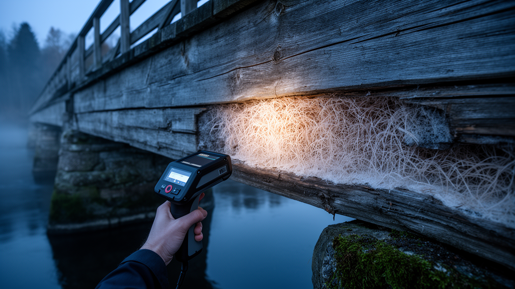
Beneath the weathered grey planks of Sweden’s oldest wooden bridge, an invisible enemy has been quietly consuming the timber from the inside out. The Lejonström bridge, a 207.5-metre structure spanning the Skellefte River in northern Sweden since 1737, has survived nearly three centuries of harsh subarctic seasons, automobile traffic, and repeated floods. Now, a team of researchers at Luleå University of Technology, working with the Swedish University of Agricultural Sciences, has combined hospital-grade imaging technology with DNA detective work to catch the culprits red-handed. Their findings, published in npj Heritage Science, reveal that even the most durable heartwood timber is no match for specialised wood-decay fungi thriving in a changing northern climate.
The research focused on timber elements removed during the bridge’s most recent renovation in the autumn of 2025, when workers from the Skellefteå Municipality replaced roadway and frame sections that showed visible signs of rot. Because the bridge has been a state-protected architectural monument since 1994 and cannot be rebuilt, moved, or fundamentally altered, conservationists face a peculiar challenge: they must monitor and maintain a living piece of history without destroying the very fabric that makes it precious. Destructive sampling is severely restricted, which means that much of the internal damage to such structures historically went undetected until it became catastrophic.
Enter X-ray computed tomography, or CT scanning, the same non-invasive technology developed for medical diagnostics that has more recently transformed wood science. The team used a Microtec Mito scanner equipped with a cone-beam X-ray source to peer inside the removed bridge timbers at a spatial resolution of half a millimetre per voxel. What they saw was striking. In three-dimensional reconstructions processed with Python and visualised in 3D Slicer, intact wood could be clearly distinguished from decayed regions by density alone. Sound wood measured approximately 600 kilograms per cubic metre, while degraded zones dropped to around 450. In one particularly damaged specimen, the heartwood around the pith had decayed so completely that a central cavity had formed, and surrounding density fell to just 330 kilograms per cubic metre against roughly 520 in adjacent sound tissue.
The CT images did more than quantify the damage; they told a story about how it developed. Density gradients along the longitudinal sections suggested that decay had initiated at one end of the timbers and progressed inward, most likely accelerated by long-term moisture exposure. The scans even revealed an unexpected accomplice: a metal nail embedded deep within one timber, whose presence may have drawn moisture into the wood through suction from the end-grain, creating a localised microenvironment ideal for fungal colonisation. Crucially, the degraded regions corresponded to areas showing the classic fingerprints of brown rot, including cubical cracking, where the wood shrinks and fractures into small, brittle, cube-like chunks as its structural polymers are dismantled.
Brown rot is among the most destructive forms of fungal decay in conifer timber. Unlike white-rot fungi, which enzymatically degrade all major cell-wall components including the tough lignin, brown-rot fungi primarily depolymerise cellulose and hemicellulose through a combination of oxidative and enzymatic mechanisms, leaving behind modified lignin residues and wood that has lost most of its mechanical strength. Because this degradation often begins deep inside a beam long before any external symptom appears, traditional visual inspection frequently fails. The density loss documented by the Swedish team demonstrates precisely why non-destructive imaging is becoming indispensable for heritage structures: externally, some of these timbers showed only limited visible damage while harboring extensive internal deterioration.
To identify which organisms were responsible, the researchers turned to culture-based microbiology. Wood fragments were separated from timber sections 50 millimetres beyond visible damage edges, surface-sterilised with sodium hypochlorite and ethanol, and placed on malt extract agar in controlled laboratory chambers. In a clever methodological refinement, additional fragments were briefly flame-sterilised to suppress fast-growing surface moulds, a step that proved essential for recovering slow-growing decay fungi that would otherwise be overrun. The team also hunted microscopically for clamp connections, distinctive loop-like structures on hyphae that betray basidiomycete identity. The result was a library of 37 pure fungal cultures representing 17 species across 11 genera, identified by amplifying and Sanger-sequencing the internal transcribed spacer (ITS) region of fungal DNA and comparing the sequences against the GenBank database, with sequences deposited under accession numbers PZ161136 through PZ161152.
Most of the recovered species were saprotrophs, moulds, and wood-associated ascomycetes typical of weathered timber, including multiple species of Trichoderma and Penicillium, along with Aureobasidium pullulans, Mucor circinelloides, Apiospora arundinis, and Sydowia polyspora. These are largely secondary colonisers, opportunists exploiting the nutrient-rich niches created as decomposition advances. Trichoderma colonies, in particular, grew so aggressively in the laboratory that they blanketed culture plates within three to four days. But the headline discovery involved two basidiomycetes found in association with the brown-rot symptoms: Amyloporia xantha, isolated seven times and the most frequently recovered basidiomycete, and a single isolate of Crustoderma sp., which could not be pinned down to species level with the ITS marker alone.
Amyloporia xantha is a well-recognised conifer-associated brown-rot fungus with a documented track record of degrading structural timber across northern Europe. Surveys in Norway have found this genus affecting roof and wall constructions, roof beams, and exterior structures, while a study of historic pre-1945 buildings in Styria, Austria, identified it in more than ten percent of cases, second only to the notorious dry-rot fungus Serpula. Its frequent recovery from the Lejonström timbers strongly suggests it is a principal driver of the bridge’s brown rot. The detection of Crustoderma adds an intriguing ecological wrinkle: many species in this genus are rare and of conservation interest, and the most frequently reported Swedish species, Crustoderma corneum, is classified as Near Threatened on the Swedish Red List. Its presence may reflect historic use of heartwood from old-growth forests rather than aggressive invasion, making the bridge something of an ecological time capsule as well as an engineering monument.
Phylogenetic analysis using Maximum Likelihood methods confirmed the picture, separating the ascomycetes, basidiomycetes, and the sole Mucoromycota representative into well-supported distinct lineages, with A. xantha and Crustoderma clustering together within the basidiomycete clade at 96 percent bootstrap support. The researchers acknowledge limitations: relying on a single culture medium likely favoured fast-growing species, and culture-independent metabarcoding could reveal slow-growing or unculturable inhabitants of historic timber. Still, the integrated approach of pairing CT-based structural assessment with molecular fungal identification offers a powerful template for heritage monitoring elsewhere.
The stakes are rising. Climate change is expected to increase temperatures, alter precipitation, and prolong periods of elevated wood moisture across northern environments, conditions that favour fungal growth and colonisation. The coastal climate of Västerbotten County, with relative humidity fluctuating between roughly 65 and 92 percent across the year, already provides ample opportunity for decay organisms. For the Lejonström bridge, which today looks much as it did in the 1870s and still carries automobile traffic, the study delivers both a warning and a toolkit: hidden decay is real, it is progressing, and X-ray tomography combined with fungal DNA sequencing can now catch it early enough to act. As the authors conclude, long-term surveillance of fungal-associated degradation will become increasingly vital for preserving wooden cultural heritage, and monitoring schedules should be tailored to each structure’s environmental exposure and conservation status rather than following a universal inspection interval.
Subject of Research: Brown-rot fungal decay and non-destructive CT monitoring of timber in the historic Lejonström bridge, Sweden
Article Title: Characterisation of brown-rot decay in the historic Lejonström bridge using X-ray tomography and molecular identification of associated fungi
Article References: Postovoitova, A., Lin, C.-F., Davydenko, K., Broman, O., Karlsson, O., & Myronycheva, O. (2026). Characterisation of brown-rot decay in the historic Lejonström bridge using X-ray tomography and molecular identification of associated fungi. npj Heritage Science, 14(1), Article 514. https://doi.org/10.1038/s40494-026-02998-3
Image Credits: AI Generated
DOI: 10.1038/s40494-026-02998-3
Keywords: brown rot, wood decay fungi, X-ray computed tomography, cultural heritage, Lejonström bridge, Amyloporia xantha, Crustoderma, ITS sequencing, Scots pine, timber conservation, mycology, Sweden
Cite Scienmag News
Roger Howard. (October 15, 2026). X-rays Reveal Hidden Fungus Eating Sweden’s Oldest Wooden Bridge. Scienmag. https://scienmag.com/x-rays-reveal-hidden-fungus-eating-swedens-oldest-wooden-bridge/
Roger Howard. "X-rays Reveal Hidden Fungus Eating Sweden’s Oldest Wooden Bridge." Scienmag, 15 October 2026, https://scienmag.com/x-rays-reveal-hidden-fungus-eating-swedens-oldest-wooden-bridge/. Accessed 15 October 2026.
Roger Howard. "X-rays Reveal Hidden Fungus Eating Sweden’s Oldest Wooden Bridge." Scienmag. October 15, 2026. https://scienmag.com/x-rays-reveal-hidden-fungus-eating-swedens-oldest-wooden-bridge/


