


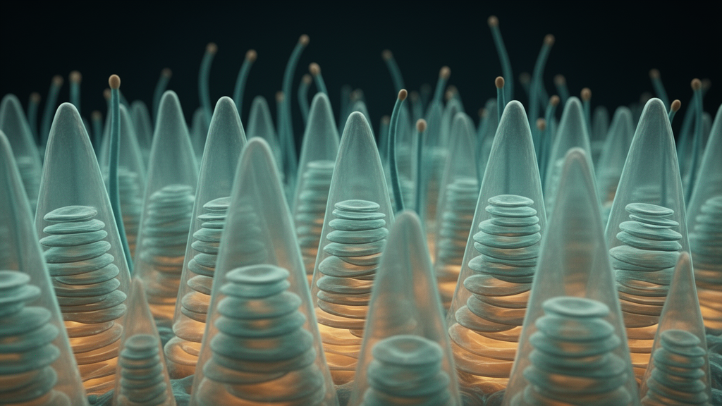
For more than six decades, vision scientists have argued about one of the most fundamental architectural questions in the eye: what exactly do the light-sensing membranes inside cone photoreceptors look like at the finest scale? Cones are the cells in the retina that make color vision and daytime sight possible, and at their tip sits a stack of flattened membrane sacs called disks, the structures that capture photons and convert them into neural signals. In non-mammalian species such as fish, amphibians, and birds, researchers have long established that these disk membranes typically remain continuous with the surrounding plasma membrane of the cell, leaving them open to the extracellular environment. Whether the same design holds in mammals, and especially in primates, has stubbornly resisted a definitive answer, because the question demands imaging and physiological measurements at a resolution and reliability that have been extraordinarily difficult to achieve in primate retinal tissue.
Now a study led by Dr. David S. Williams, professor of ophthalmology and neurobiology at the David Geffen School of Medicine at UCLA, in collaboration with researchers at the University of California, Santa Barbara and Harvard, reports that the answer for primates is the opposite of what is seen in many other vertebrates. Working with retinal tissue near the fovea of Rhesus monkeys (Macaca mulatta), the team found that the majority of disk membranes in primate cones are closed, self-contained structures, much like the disks found in rod photoreceptors, the cells responsible for vision in dim light. The work, published in the Journal of Neuroscience, represents the first quantitative assessment of the relative proportion of open and closed disk membranes in a mammalian cone, and it settles a debate that has lingered since the earliest electron micrographs of the retina were first interpreted.
The significance of the question is easier to appreciate when one considers what the disks actually do. Phototransduction, the molecular cascade that turns a single photon into an electrical signal, takes place on these membranes. Light-sensitive opsin proteins and the G protein transducin are embedded in the disk membrane, and the second messenger cyclic GMP, along with calcium ions, diffuses in the compartments defined by the disk geometry. Whether the interior of a disk communicates freely with the space outside the cell or is sealed off changes the diffusion pathways available to these signaling molecules, the surface area available for protein trafficking, and the way in which the cell renews its light-sensitive machinery. Structure, in other words, is not a cosmetic detail; it constrains the entire operating logic of the photoreceptor.
Historically, the debate arose because different experimental approaches yielded apparently conflicting pictures. Early transmission electron microscopy of retinal sections produced images that some investigators interpreted as showing continuity between the disk rim and the plasma membrane, while others saw sealed, independent sacs. Preparation artifacts, the extreme fragility of photoreceptor outer segments, and the sheer difficulty of cutting and imaging tissue thin enough to resolve the relevant membranes all conspired to keep the question open. In non-mammalian cones, serial sectioning and physiological measurements converged on the conclusion that the disks are generally open, continuous evaginations of the plasma membrane, and many researchers assumed that mammalian cones would follow the same plan. Without quantitative data from primate tissue, however, the assumption remained exactly that: an assumption.
The UCLA-led team attacked the problem with two independent methods, each capable of addressing the question from a different direction. The first was electron tomography, a technique that goes beyond conventional two-dimensional electron micrographs by reconstructing three-dimensional volumes of tissue from a series of images taken at different tilt angles. Rather than inferring membrane topology from a single thin slice, tomography allows investigators to trace membranes through the volume and determine whether a given disk is sealed or whether it opens onto the extracellular space. Applied to the cone outer segments of the foveal region in Rhesus monkey retina, this approach provided direct structural evidence about the patency, or openness, of individual disk membranes.
The second approach was electrophysiological. If cone disks were open to the extracellular space, the electrical properties of the cell would reflect that continuity in measurable ways, because the membrane area accessible to ion channels and to the diffusion of charged signaling molecules would be organized differently than in a cell with sealed disks. By recording from primate cone photoreceptors and analyzing the electrical signatures of the outer segment, the researchers obtained functional evidence that could be compared directly against the structural data. The power of the study lies in this convergence: two methods with entirely different sources of potential error pointed to the same conclusion, and that conclusion contradicted the pattern established in non-mammalian vertebrates.
Through both approaches, the researchers found that the majority of disk membranes in primate cones are closed, independent structures. This is the configuration familiar from rod photoreceptors, whose disks are well established to be enclosed sacs that are periodically shed at the tip of the outer segment and renewed through new membrane synthesis at the base. The finding means that primate cones, despite their distinct morphology and their role in high-acuity color vision, share with rods the fundamental design principle of sealed disk membranes. It also means that textbooks and models of cone function, many of which have implicitly carried the open-disk assumption into discussions of mammalian and human vision, will need to be revised.
The implications extend into several areas of cell biology and physiology. Disk membrane renewal is one of the most remarkable housekeeping operations in the body: photoreceptors constantly rebuild their light-sensitive stacks, and in rods this involves the daily shedding and phagocytosis of aged disk packets by the retinal pigment epithelium. Whether cones renew their disks through a comparable process has been debated in its own right, and the closed architecture documented in this study provides a structural framework for understanding how such renewal could proceed in primate cones. Similarly, models of phototransduction depend on assumptions about the compartments in which cyclic GMP and calcium diffuse; sealed disks create a distinct intradiskal space whose ionic composition can be regulated separately from the extracellular environment, altering the dynamics of adaptation, the process by which photoreceptors adjust their sensitivity across enormous ranges of light intensity.
Because the tissue examined came from the region near the fovea, the part of the primate retina responsible for our sharpest, most detailed vision, the findings speak directly to human visual physiology. The Rhesus macaque is a well-established model for human retinal structure and function, and the cone-dominant foveal region is precisely where the stakes of the structural question are highest. Understanding that the disks in these cells are closed brings the structural biology of primate cones into alignment with the electrophysiological models that have been refined over decades, and it offers a corrected foundation for studying retinal diseases that target cones, including the macular degenerations that rob millions of people of central vision. The authors declare no competing financial interests, and the research was published as a peer-reviewed contribution to the Journal of Neuroscience under the title describing the ultrastructural and electrophysiological determination of monkey cone outer segment membrane patency.
What makes the study a striking example of basic science at its best is that it resolves, with modern tools, a question posed at the dawn of retinal ultrastructure. The combination of three-dimensional electron tomography with complementary electrophysiology provided the quantitative rigor that earlier generations of microscopists could not achieve, and the answer it delivered, that primate cone disks are closed like those of rods, reshapes how scientists will model phototransduction, disk renewal, and cone physiology in humans. After sixty years of uncertainty, the architecture of the cells that give primates their remarkable color vision is finally clear, and that clarity opens the door to a deeper understanding of how the eye builds sight from stacks of membrane only nanometers thick.
Subject of Research: Structural patency of disk membranes in primate cone photoreceptors
Article Title: Study from the Stein Eye Institute answers long-debated structural question of primate cone photoreceptors
Article References: Study from the Stein Eye Institute answers long-debated structural question of primate cone photoreceptors. (n.d.). Original publication
Image Credits: AI Generated
DOI: Not provided
Keywords: cone photoreceptors, retina, disk membranes, electron tomography, electrophysiology, phototransduction, Rhesus macaque, fovea, vision science, UCLA, Journal of Neuroscience, photoreceptor structure
Cite Scienmag News
Cassandra Pierce. (October 9, 2026). Primate Cone Cells Solved: Light-Sensing Disks Are Closed After Decades of Debate. Scienmag. https://scienmag.com/primate-cone-cells-solved-light-sensing-disks-are-closed-after-decades-of-debate/
Cassandra Pierce. "Primate Cone Cells Solved: Light-Sensing Disks Are Closed After Decades of Debate." Scienmag, 9 October 2026, https://scienmag.com/primate-cone-cells-solved-light-sensing-disks-are-closed-after-decades-of-debate/. Accessed 9 October 2026.
Cassandra Pierce. "Primate Cone Cells Solved: Light-Sensing Disks Are Closed After Decades of Debate." Scienmag. October 9, 2026. https://scienmag.com/primate-cone-cells-solved-light-sensing-disks-are-closed-after-decades-of-debate/


