


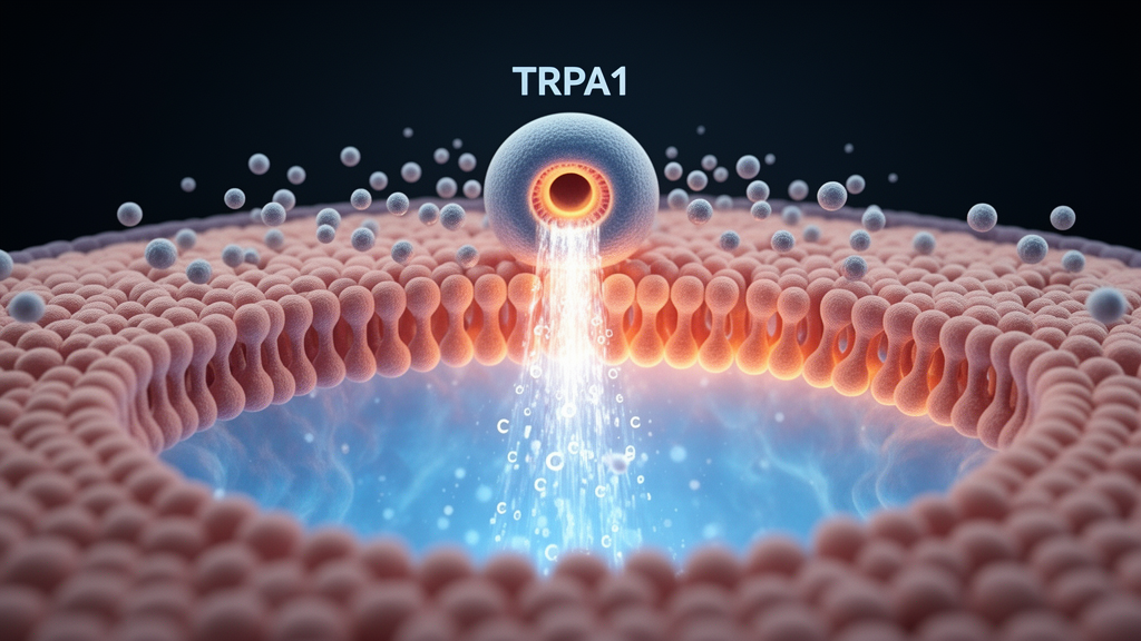
Lipid nanoparticles have become one of the most celebrated delivery vehicles in modern medicine, ferrying messenger RNA into cells for vaccines and a growing list of experimental therapies. Yet a new study suggests that these synthetic fat droplets do far more than quietly deposit their cargo. According to research published in Cellular and Molecular Life Sciences by Alina Milici, Justyna B. Startek, Geert Bultynck and Karel Talavera of KU Leuven, lipid nanoparticles can provoke a striking burst of calcium signaling inside cells, and a well-known pain-sensing ion channel called TRPA1 is a key player in that response. The finding adds an unexpected dimension to how scientists think about the immediate interactions between these nanoparticles and the cells they are meant to treat.
TRPA1 is what biologists call a polymodal ion channel receptor, meaning it can be switched on by an unusually wide range of stimuli. It sits in the plasma membrane of nerve endings and many other cell types, and it is best known for its role in nociception, the neural process that produces the sensation of pain. Mustard oil, allicin from garlic, irritants in smoke and even compounds produced during tissue injury can all activate TRPA1, which then opens a pore that lets positively charged ions, including calcium, flood into the cell. That calcium influx is the electrical and chemical signal that ultimately alerts the nervous system.
What makes the new work particularly intriguing is the mechanism the Belgian team proposes. TRPA1, they note, can be activated by local mechanical perturbations in the surrounding plasma membrane, specifically by molecules that physically insert themselves into the lipid bilayer. Lipid nanoparticles are, in essence, small blobs of engineered lipid that must fuse with or otherwise engage the plasma membrane to deliver their contents. The researchers therefore asked a deceptively simple question: when lipid nanoparticles touch a target cell, do they perturb the membrane in a way that modulates TRPA1 function? Their answer was yes, and the consequences turned out to be more complex than a single channel opening.
Using cells engineered to express TRPA1 as well as native cells that carry the channel naturally, the team observed that exposure to lipid nanoparticles produced irregular calcium transients, meaning rapid, fluctuating rises in the concentration of calcium ions in the cytosol. The erratic, unpredictable character of these signals is telling. Rather than a clean, uniform response, the researchers interpret the pattern as reflecting stochastic interactions between individual nanoparticles and the plasma membrane. Each encounter between a nanoparticle and the cell surface is a chance event, and the sum of many such events produces a noisy, irregular calcium signature rather than a synchronized wave.
To dissect what was actually happening, the investigators applied both selective and non-selective TRPA1 inhibitors to their preparations. This pharmacological dissection revealed that the cytosolic calcium transients arise through several distinct mechanisms operating in parallel. Some of the calcium enters the cell through TRPA1-dependent influx, confirming that the channel genuinely contributes to the nanoparticle response. But a substantial component of the signal persists even when TRPA1 is blocked, indicating a TRPA1-independent calcium influx pathway that the nanoparticles can engage. On top of both influx routes, the team found evidence of calcium being mobilized from internal stores, specifically released from the endoplasmic reticulum, the cell’s main calcium reservoir. The nanoparticle effect, in other words, is not a single event but a layered cascade touching multiple arms of cellular calcium handling.
One of the most consequential discoveries involves acidity. The researchers found that the responses to lipid nanoparticles were enhanced when the extracellular environment was made more acidic, that is, when the pH was lowered. Probing further, they determined that low pH increases the effects of the nanoparticles on membrane fluidity, the ease with which lipid molecules in the bilayer move and rearrange. A more fluid membrane is presumably more susceptible to the perturbations that nanoparticles impose, and that susceptibility translates into stronger calcium signaling. This detail matters enormously for medicine, because many of the tissues where lipid nanoparticle therapies are deployed, including inflamed tissues and certain tumors, are naturally acidic. A vaccine or drug delivered by lipid nanoparticles into an acidic microenvironment may therefore provoke a different and stronger cellular response than the same formulation in neutral conditions.
The authors are careful to frame their results as unveiling a non-canonical activation mechanism for TRPA1. Classical TRPA1 agonists are reactive chemicals that covalently modify cysteine residues on the channel, but membrane-inserting agents represent a fundamentally different way of gating the pore. Synthetic lipid nanoparticles now join the roster of agents capable of engaging the channel through bilayer perturbation, a category that had previously been populated mainly by amphipathic molecules and certain local anesthetics. For the ion channel field, this expands the conceptual map of how mechanical and lipid-based forces can be transduced into electrical and calcium signals.
For the drug delivery field, the implications are equally significant. The acute effects of lipid nanoparticles on the plasma membrane and the signaling pathways coupled to it had remained poorly characterized, even as these particles became the backbone of mRNA vaccine technology during the COVID-19 pandemic and are now being explored for gene therapy, cancer immunotherapy and personalized medicine. If lipid nanoparticles routinely trigger calcium influx, endoplasmic reticulum calcium release and TRPA1 activation on contact with target cells, those events could influence how cells respond to the delivered cargo, potentially shaping the potency and reactogenicity of LNP-based formulations. The authors explicitly note that these previously unrecognized effects on intracellular calcium signaling may be relevant for the development of LNP-based vaccines, where innate immune activation is a double-edged sword: enough to provoke immunity, but not so much as to cause harmful inflammation.
There is also a sensory dimension worth considering. TRPA1-expressing nerve endings are the sentinels that detect chemical irritation and pain throughout the body. If circulating or injected lipid nanoparticles can activate this channel, it raises the possibility that some of the acute side effects associated with nanoparticle administration, from injection-site discomfort to broader inflammatory sensations, could involve direct sensory channel activation rather than downstream immune mediators alone. The study did not set out to test pain in living organisms, so such connections remain hypotheses for future work, but the mechanistic groundwork is now in place.
Methodologically, the study exemplifies the power of combining heterologous expression systems, native cells and pharmacology to untangle a multi-component signal. The CHO cell line expressing mouse TRPA1, kindly provided by Ardem Patapoutian’s laboratory at The Scripps Research Institute, allowed the team to isolate channel-specific effects, while native TRPA1-expressing cells confirmed that the phenomenon is not an artifact of overexpression. Calcium imaging served as the readout, capturing the dynamic interplay between plasma membrane influx and endoplasmic reticulum release in real time. The work was supported by the Research Foundation Flanders, or FWO, and the authors are partners of the FWO Scientific Research Network CaSign, reflecting Belgium’s strong tradition in calcium signaling research. As lipid nanoparticles continue their march from the clinic into new therapeutic frontiers, this study is a reminder that the delivery vehicle itself is an active biological agent, capable of switching on one of the body’s most sensitive molecular alarms the moment it touches a cell.
Subject of Research: Activation of the TRPA1 ion channel and calcium signaling by synthetic lipid nanoparticles
Article Title: TRPA1 contributes to calcium signaling induced by synthetic lipid nanoparticles
Article References: Milici, A., Startek, J. B., Bultynck, G., & Talavera, K. (2026). TRPA1 contributes to calcium signaling induced by synthetic lipid nanoparticles. Cellular and Molecular Life Sciences. https://doi.org/10.1007/s00018-026-06476-8
Image Credits: AI Generated
DOI: 10.1007/s00018-026-06476-8
Keywords: TRPA1, lipid nanoparticles, calcium signaling, ion channels, plasma membrane, endoplasmic reticulum, membrane fluidity, low pH, nociception, mRNA vaccines, drug delivery, calcium imaging
Cite Scienmag News
Drew Townsend. (October 9, 2026). Lipid Nanoparticles Trigger Pain Receptor TRPA1 and Reshape Calcium Signaling in Cells. Scienmag. https://scienmag.com/lipid-nanoparticles-trigger-pain-receptor-trpa1-and-reshape-calcium-signaling-in-cells/
Drew Townsend. "Lipid Nanoparticles Trigger Pain Receptor TRPA1 and Reshape Calcium Signaling in Cells." Scienmag, 9 October 2026, https://scienmag.com/lipid-nanoparticles-trigger-pain-receptor-trpa1-and-reshape-calcium-signaling-in-cells/. Accessed 9 October 2026.
Drew Townsend. "Lipid Nanoparticles Trigger Pain Receptor TRPA1 and Reshape Calcium Signaling in Cells." Scienmag. October 9, 2026. https://scienmag.com/lipid-nanoparticles-trigger-pain-receptor-trpa1-and-reshape-calcium-signaling-in-cells/


