


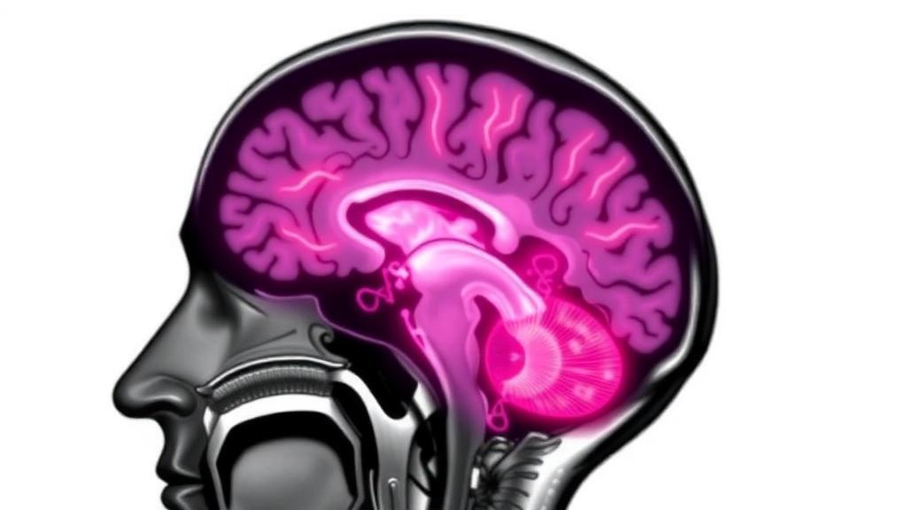
It begins, in most cases, with something deceptively mundane: a patch of numbness on one side of the face. Over the following years, that numbness spreads across the scalp and into the limbs, while muscles of the jaw, tongue, and hands begin to waste away. This is facial onset sensory and motor neuronopathy, or FOSMN, a neurological disorder so rare that only around one hundred cases have been documented worldwide since it was first described in 2006. Because routine brain scans of affected patients almost always look normal, clinicians have long lacked objective evidence of what the disease does inside the brain. Now a team at University Hospital Ulm in Germany has applied advanced magnetic resonance imaging techniques to a group of six patients, and for the first time mapped a consistent neuroanatomical signature of the condition, publishing their findings in the Journal of Neurology.
The clinical picture of FOSMN is as distinctive as it is devastating. Patients typically notice unilateral facial tingling or numbness, followed by progressive weakness that advances over a span of two to six years, eventually encompassing the entire face, scalp, and upper limbs. Bulbar symptoms, including difficulty swallowing and speaking, often emerge within six years of onset. A hallmark feature is atrophy of the temporal and masseter muscles, the muscles of chewing, which can give the face a hollowed appearance. Both sensory and motor deficits follow a strict rostro-caudal trajectory, marching downward from the head toward the hands, with profound muscle wasting documented as late as ten years after the first symptoms. Lower motor neuron signs, particularly muscle atrophy, are present in every documented case.
Diagnosis has traditionally rested on electrophysiology rather than imaging. A prolonged or absent blink reflex is a key clinical hallmark, while needle electromyography reveals neurogenic denervation and reinnervation across cranial, cervical, and thoracic levels. Sensory nerve conduction studies typically show reduced amplitudes in the upper limbs. Conventional brain MRI, cerebrospinal fluid analysis, and laboratory work are generally unremarkable, although scans may occasionally show atrophy of the tongue or spinal cord. The main differential diagnoses include bulbar-onset amyotrophic lateral sclerosis, which is purely motor and lacks the facial sensory loss characteristic of FOSMN, as well as Bickerstaff brainstem encephalitis and Miller Fisher syndrome, which are distinguished by their acute, postinfectious course, eye movement palsies, and ataxia, in sharp contrast to the slow neurodegenerative progression of FOSMN.
What causes FOSMN remains unknown, but growing evidence points to an intriguing overlap with two better-known neurodegenerative conditions: amyotrophic lateral sclerosis and frontotemporal dementia. The motor features of FOSMN, with bulbar weakness and muscle atrophy, resemble ALS, while a subset of patients develops cognitive deficits and behavioral changes reminiscent of frontotemporal dementia. Genetic studies have strengthened this connection, identifying mutations in ALS-associated genes including SOD1, TARDBP, and SQSTM1 in some patients. Postmortem examinations have revealed significant atrophy of the trigeminal nerve roots, degenerative changes in both the sensory and motor nuclei of the trigeminal nerve, and substantial loss of anterior horn cells, the spinal motor neurons that control voluntary movement. Yet no accepted diagnostic criteria exist, and the fundamental question of whether FOSMN is neurodegenerative or immune-mediated remains open.
To probe the disease in living patients, the Ulm team, led by Angela Rosenbohm with Hans-Peter Müller and Jan Kassubek as senior authors, recruited six consecutive patients with clinically confirmed FOSMN, four men and two women aged 48 to 84 years, who presented to their department between 2016 and 2026. Given that only about one hundred cases exist worldwide, even a group of six represents a substantial cohort for this condition. The patients were compared with thirty age- and sex-matched healthy controls, all free of neurological, psychiatric, or major medical illness and without gross brain pathology on conventional MRI. The study was approved by the Ethics Committee of the University of Ulm and conducted in accordance with the Declaration of Helsinki, with written informed consent from all participants.
The researchers deployed two complementary, unbiased analysis techniques on data acquired from a 1.5 Tesla scanner. The first, atlas-based volumetry, processed T1-weighted structural images by segmenting them into gray matter, white matter, and cerebrospinal fluid compartments, normalizing them into standard Montreal Neurological Institute space, and measuring volumes of predefined brain regions using the LONI Probabilistic Brain Atlas. All results were standardized to the mean intracranial volume of the controls. The second technique, diffusion tensor imaging combined with whole brain-based spatial statistics, probed the microstructure of white matter tracts by measuring fractional anisotropy, a metric that reflects the directional organization of water diffusion and therefore the integrity of nerve fiber bundles. Statistical comparisons used the Welch test with false discovery rate correction and a cluster-size threshold to guard against spurious findings.
The volumetric results were striking. At the group level, FOSMN patients showed significant global brain atrophy, but the most pronounced volume loss was localized to the medulla oblongata, the lowermost portion of the brainstem that houses the nuclei of the trigeminal nerve and the medial lemniscus, a major sensory pathway. The insula, a deep cortical region involved in integrating bodily sensations, showed the next highest degree of shrinkage. Across the cerebral lobes, gray matter was generally more affected than white matter. This combination of widespread cerebral volume loss with a focal brainstem emphasis is anatomically plausible for a disorder that attacks the cranial nerves governing facial sensation and the bulbar muscles controlling speech and swallowing.
The diffusion imaging results added a second layer of insight. Fractional anisotropy was significantly reduced in the right frontal lobe and, bilaterally, along the superior and inferior longitudinal fasciculi, two major association pathways. The superior longitudinal fasciculus forms a large arcuate bundle connecting the frontal lobes with the parietal and temporal regions, while the inferior longitudinal fasciculus is an occipitotemporal projection system. Notably, the corticospinal tract, the primary motor pathway whose degeneration is the classic imaging signature of ALS, showed no significant involvement. The affected tracts are polymodal integration highways, and their disruption mirrors patterns previously described in frontotemporal lobar degeneration, aligning with the clinical and genetic overlap between FOSMN and that spectrum of disease. Interestingly, while direct connections to the face area of the somatosensory cortex did not fall within the significant clusters, trigeminal nerve disorders such as trigeminal neuralgia have previously been linked to microstructural alterations along the superior longitudinal fasciculus.
The authors are careful about the limitations. Six patients cannot settle every question, and diffusion tensor imaging cannot establish the directionality of tract changes, so the findings do not explain the characteristic rostro-caudal march of symptoms. Only one patient in the cohort showed upper motor neuron signs, so corticospinal tract involvement in more motor-dominant cases remains to be tested. Nor can MRI resolve whether the underlying process is neurodegenerative or immune-mediated. Yet the consistency of the volumetric and microstructural findings, obtained with standardized protocols on a single scanner, constitutes a genuine advance: for the first time, routine-looking scans have yielded an objective, in vivo neuroanatomical pattern for a disease whose biology has been almost entirely inferred from case reports and autopsies.
The clinical profiles of the six patients underscore the severity of the condition. Three presented with facial numbness, one with oropharyngeal dysphagia, one with weakness of the masticatory muscles, and one with forearm weakness. Swallowing impairment was confirmed by fiberoptic endoscopic evaluation in five patients, and two required feeding tubes placed directly into the stomach. Two patients developed upper-limb paresis a mean of 48 months after diagnosis, with intervals ranging from 12 to 84 months. The mean follow-up was just over four years. The team emphasizes that future progress will require multicenter studies with standardized clinical and neuroimaging assessments, ideally incorporating spinal cord imaging and longitudinal measurements that track how the brain changes as individual symptoms progress. Such phenotype-neuroimaging correlations, they argue, are the key to finally understanding what drives this enigmatic disorder, and potentially to distinguishing it earlier from the ALS-like and inflammatory conditions it so often mimics.
Subject of Research: Neuroimaging signatures of facial onset sensory and motor neuronopathy (FOSMN)
Article Title: Neuroimaging signatures of facial onset sensory and motor neuronopathy (FOSMN)
Article References: Rosenbohm, A., Herrera-Angeles, F., Huppertz, H.-J., Haeusler, K. G., Weishaupt, J. H., Müller, H.-P., & Kassubek, J. (2026). Neuroimaging signatures of facial onset sensory and motor neuronopathy (FOSMN). Journal of Neurology, 273(10), Article 639. https://doi.org/10.1007/s00415-026-14174-5
Image Credits: AI Generated
DOI: 10.1007/s00415-026-14174-5
Keywords: FOSMN, neuroimaging, diffusion tensor imaging, atlas-based volumetry, motor neuron disease, amyotrophic lateral sclerosis, frontotemporal dementia, medulla oblongata, white matter tracts, brainstem, rare disease, MRI
Cite Scienmag News
Cassandra Pierce. (October 5, 2026). Rare Facial-Onset Nerve Disease Reveals Its Brain Footprint in Advanced MRI Study. Scienmag. https://scienmag.com/rare-facial-onset-nerve-disease-reveals-its-brain-footprint-in-advanced-mri-study/
Cassandra Pierce. "Rare Facial-Onset Nerve Disease Reveals Its Brain Footprint in Advanced MRI Study." Scienmag, 5 October 2026, https://scienmag.com/rare-facial-onset-nerve-disease-reveals-its-brain-footprint-in-advanced-mri-study/. Accessed 5 October 2026.
Cassandra Pierce. "Rare Facial-Onset Nerve Disease Reveals Its Brain Footprint in Advanced MRI Study." Scienmag. October 5, 2026. https://scienmag.com/rare-facial-onset-nerve-disease-reveals-its-brain-footprint-in-advanced-mri-study/


