


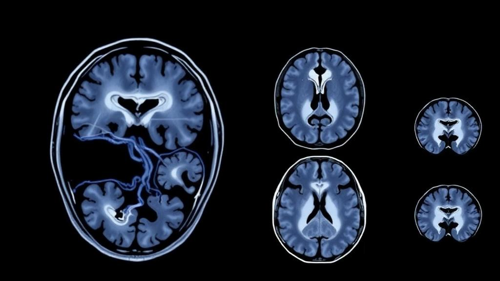
Every year, thousands of children around the world are diagnosed with high-grade gliomas, aggressive tumors of the brain and spinal cord that remain among the leading causes of cancer-related death in childhood. For families, one of the most agonizing uncertainties is prognosis: even after surgery, radiation, and chemotherapy, doctors often struggle to say with confidence how a particular child is likely to fare. A new multi-institution study published in the Journal of Neuro-Oncology suggests that a powerful part of the answer may already be hiding in plain sight, inside the routine magnetic resonance imaging scans that every one of these patients receives at diagnosis.
The research, led by Mariam Tolba and Michael Zhang of Stanford University School of Medicine together with senior authors Laura M. Prolo and Kristen W. Yeom, harnessed a computational technique known as radiomics. Rather than relying on what a radiologist can see with the naked eye, radiomics converts medical images into vast troves of quantitative data. The team extracted 1,800 standardized features from gadolinium-enhanced T1-weighted and T2-weighted MRI scans of 77 children with non-midline, hemispheric high-grade gliomas, tumors that arise in the brain’s hemispheres rather than along its central structures. Every feature followed the Image Biomarker Standardisation Initiative, an international framework designed to make image-derived measurements reproducible across scanners, hospitals, and countries.
The children in the cohort, whose average age was 140 months and who included 43 males, were treated at five different pediatric institutions, a deliberate design choice that reflects the real-world diversity of MRI machines, imaging protocols, and patient populations. All scans were treatment-naïve, meaning they were acquired before any surgery, radiation, or chemotherapy had altered the tumor’s appearance. This detail matters enormously: the goal was to capture the tumor’s intrinsic biology at the moment of diagnosis, not the artifacts of treatment. From each tumor, the pipeline measured characteristics such as shape, texture, intensity distributions, and the fine-grained spatial patterns that describe how heterogeneously a tumor enhances with contrast, properties that often correlate with underlying features like cell density, necrosis, and blood-brain barrier disruption.
To translate this feature library into a clinically useful prediction, the researchers turned to Cox proportional hazards regression, a statistical framework built specifically for modeling survival outcomes. Using k-fold cross-validation, a technique that repeatedly trains the model on subsets of the data and tests it on held-out patients, they identified the optimal combination of features for predicting overall survival. Each patient then received a risk score, calculated as a linear combination of the selected features weighted by their regression coefficients. Splitting the cohort at the median risk score produced two groups: a high-risk group and a low-risk group. All model development was performed in Python, and in a move that will please the open-science community, the complete analysis code has been released on GitHub and archived on Zenodo.
The results are striking. A model combining clinical variables, specifically age and sex, with MRI-derived radiomics features achieved a concordance index of 0.78, with a 95 percent confidence interval of 0.70 to 0.83. The concordance index, sometimes called the C-index, measures how well a model ranks patients by risk; a value of 0.5 is no better than a coin flip, while 1.0 represents perfect discrimination. Radiomics features alone reached 0.75, whereas clinical features alone managed only 0.61. In other words, the quantitative texture and shape information buried in the scans carried substantially more prognostic signal than basic demographic data, and combining the two sources produced the best performance of all.
The survival gap between the risk groups was clinically meaningful. Children classified as high risk by the radiomics model had a median overall survival of 21.7 months, while those in the low-risk group survived a median of 44.6 months, more than twice as long. The difference was statistically robust, with a log-rank P value of 0.007 and a hazard ratio of 2.42, meaning children in the high-risk group faced roughly two and a half times the risk of death at any given time compared with their low-risk counterparts, with a 95 percent confidence interval of 1.26 to 4.66. When visualized as Kaplan-Meier curves, the two groups separated cleanly, offering an intuitive picture of how a mathematical score derived from pixels maps onto the lived trajectories of young patients.
What makes this approach so compelling is that pediatric high-grade gliomas are biologically distinct from their adult counterparts. The 2021 World Health Organization classification of central nervous system tumors reorganized glioma diagnostics around molecular genetics, and it is now clear that childhood hemispheric gliomas harbor driver mutations and molecular alterations that differ from those seen in adult glioblastoma. Yet obtaining molecular profiles requires tissue, typically from surgery or biopsy, which carries risk and is not always feasible. Radiomics offers a non-invasive window into tumor phenotype, potentially capturing biological information that complements, and in some cases anticipates, what molecular testing reveals. Prior work by some of the same investigators has shown that similar image-based signatures can distinguish molecular subgroups of medulloblastoma, ependymoma, and diffuse intrinsic pontine glioma, suggesting this is part of a broader program to decode pediatric brain tumors through computational imaging.
The study’s authors are careful to frame the work as a pilot, and the caveats deserve attention. Seventy-seven patients, while respectable for a rare pediatric cancer, is a modest sample size, and the model will need external validation in independent, ideally larger and international, cohorts before it can influence clinical decisions. Radiomics models are also notoriously sensitive to variations in scanner manufacturer, acquisition parameters, and segmentation methods, challenges the field is actively addressing through harmonization standards like the IBSI framework the authors adopted. Still, the multi-center design, the standardized feature set, the cross-validated methodology, and the openly shared code all strengthen confidence that the findings are not an artifact of a single institution’s data.
The potential applications extend well beyond individual prognosis. In pediatric neuro-oncology, clinical trials often struggle to enroll enough patients, and every participant represents a precious resource. A validated imaging-based risk stratification tool could help refine trial eligibility, ensuring that experimental therapies are matched to the children most likely to benefit, or allowing risk-adapted treatment intensification for those flagged as high risk at diagnosis. The authors suggest that computational MRI techniques may ultimately serve a role in therapy planning, offering clinicians a quantitative, repeatable measure of tumor phenotype that can be tracked over time as treatment proceeds.
For now, the message is one of cautious optimism. The scans that children with brain tumors already undergo, at no additional cost, discomfort, or radiation exposure, contain quantifiable information that predicts survival with meaningful accuracy. As radiomics pipelines mature, become standardized across institutions, and are integrated with molecular diagnostics, the humble diagnostic MRI may evolve into something far more powerful: a computational biopsy that helps clinicians see not just where a tumor is, but what it is likely to do next. For a disease where every month of survival matters, that transformation cannot come soon enough.
Subject of Research: MRI-based radiomics for prognostic risk stratification of pediatric non-midline high-grade gliomas
Article Title: MRI-based radiomics for prognosis of non-midline, pediatric high-grade gliomas
Article References: Tolba, M., Zhang, M., Duh, J. K., Liverani, L., Chang, J., Supakul, N., Lober, R. M., Cheshier, S. H., Mattonen, S. A., Jaju, A., Prolo, L. M., & Yeom, K. W. (2026). MRI-based radiomics for prognosis of non-midline, pediatric high-grade gliomas. Journal of Neuro-Oncology, 179(3), Article 84. https://doi.org/10.1007/s11060-026-05785-z
Image Credits: AI Generated
DOI: 10.1007/s11060-026-05785-z
Keywords: radiomics, pediatric high-grade glioma, MRI, prognosis, machine learning, Cox regression, risk stratification, brain tumors, biomarkers, survival prediction, neuro-oncology, computational imaging
Cite Scienmag News
Nathaniel Bowman. (October 2, 2026). AI Reads Routine MRI Scans to Predict Survival in Children with Brain Cancer. Scienmag. https://scienmag.com/ai-reads-routine-mri-scans-to-predict-survival-in-children-with-brain-cancer/
Nathaniel Bowman. "AI Reads Routine MRI Scans to Predict Survival in Children with Brain Cancer." Scienmag, 2 October 2026, https://scienmag.com/ai-reads-routine-mri-scans-to-predict-survival-in-children-with-brain-cancer/. Accessed 2 October 2026.
Nathaniel Bowman. "AI Reads Routine MRI Scans to Predict Survival in Children with Brain Cancer." Scienmag. October 2, 2026. https://scienmag.com/ai-reads-routine-mri-scans-to-predict-survival-in-children-with-brain-cancer/


