


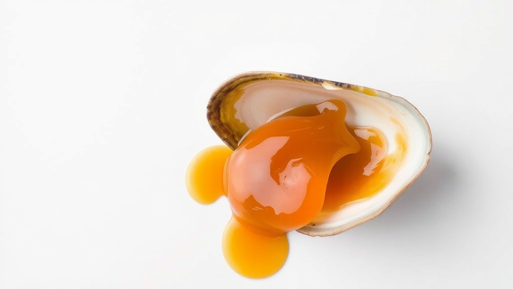
The rich brown hue of colas, sauces, beers and countless processed foods comes from caramel colourant, one of the most widely produced food additives on the planet. Yet while regulators have scrutinised these dyes mainly through the lens of human dietary safety, almost nothing has been known about what happens when they end up in seawater. A new laboratory study has now exposed a widely used food colourant class to one of marine ecotoxicology’s favourite sentinel animals, the Mediterranean mussel Mytilus galloprovincialis, and found that ammonia-based caramel colourants trigger measurable tissue damage and oxidative stress at concentrations far below what most people would guess a simple food dye could achieve.
The research, published in the journal Ecotoxicology, was carried out by a team at Çanakkale Onsekiz Mart University in Türkiye led by Emircan Aksoy and corresponding author Selin Ertürk Gürkan. The investigators focused on two of the four internationally recognised caramel colour classes: Ammonia Caramel, known as Caramel III, and Sulfite-Ammonia Caramel, known as Caramel IV. Both are produced by heating carbohydrates with ammonia-containing reagents, and both are chemically far more complex than the word caramel might suggest, containing a heterogeneous mixture of polymeric colour bodies and, in some cases, nitrogen-containing by-products such as 4-methylimidazole that have drawn toxicological attention in the past.
What makes the study unusual is its exposure route. Safety assessments of caramel colourants have overwhelmingly assumed that humans encounter them in food and drink, so comparative data on the two ammonia-based classes under waterborne conditions have been scarce. To fill that gap, the researchers exposed mussels for 96 hours to three concentrations of each caramel class: 0.05, 0.5 and 5 milligrams per litre. The top concentration was anchored to available ecotoxicological information for Caramel IV, while the lower doses allowed a controlled comparison of Caramel III under identical aquatic conditions. Mussels, which filter enormous volumes of water and are notoriously sensitive to dissolved contaminants, are considered ideal sentinels for this kind of experiment because their responses often foreshadow broader ecosystem effects.
After the exposure period, the team examined two organs with very different jobs: the gills, which handle respiration and particle capture, and the digestive gland, the mussel’s equivalent of a liver and primary site of detoxification. Under the microscope, the differences between exposed and control animals were striking. In gill tissue, the caramel treatments produced epithelial deformation, hyperplasia, an abnormal thickening of the tissue lining, fusion of the delicate gill filaments known as lamellae, infiltration by hemocytes, the molluscan immune cells, and accumulation of lipofuscin, a pigment that marks cellular wear and oxidative damage. Each of these changes can impair the gill’s ability to exchange gases and filter food, and filament fusion in particular reduces the effective surface area available for respiration.
The digestive gland told a parallel but distinct story. There, the researchers documented progressive tubular disorganisation, dilation of the tubule lumina, vacuolisation of the digestive cells and eventual epithelial atrophy. In a healthy mussel, the digestive gland tubules are tightly organised structures where intracellular digestion takes place; when they unravel and their lining cells swell with vacuoles and then waste away, the animal’s capacity to process nutrients and detoxify harmful compounds is directly compromised. The pattern of damage intensified with concentration in both organs, and the two caramel classes were not interchangeable in their effects.
Indeed, one of the study’s clearest findings was that Caramel III, the plain ammonia caramel, generally provoked stronger structural and biochemical alterations than Caramel IV at comparable concentrations. That result is notable because Caramel IV, the sulfite-ammonia variant used in dark soft drinks, has historically received more regulatory and public attention, partly because of its 4-methylimidazole content. The new data suggest that the less-studied Caramel III deserves ecotoxicological scrutiny in its own right, and the authors frame their work as the first ecotoxicological evidence for this class in a marine invertebrate model.
Structural damage was only half the picture. The team also measured a battery of oxidative stress biomarkers in both tissues, focusing on three antioxidant enzymes: superoxide dismutase, or SOD, which converts the superoxide radical into hydrogen peroxide; catalase, or CAT, which breaks hydrogen peroxide down into water; and glutathione S-transferase, or GST, a versatile detoxification enzyme that conjugates reactive electrophilic compounds to glutathione. Alongside enzyme activities, the researchers quantified lipid peroxidation, the oxidative degradation of membrane lipids that serves as a classic fingerprint of free-radical damage to cells.
The biochemical data revealed a familiar but sobering dynamic. Antioxidant enzymes were activated in parallel with rising lipid peroxidation, meaning the mussels’ cellular defence systems did respond to the caramel-induced oxidative challenge, but the compensatory response was insufficient to fully prevent oxidative damage. In other words, the animals were fighting back biochemically and still losing ground. Multivariate statistical analyses of the combined biomarker dataset confirmed that the shifts were integrated and coherent rather than random noise, producing a clear separation between control and exposed groups and reinforcing the conclusion that both caramel classes impose genuine physiological stress rather than merely cosmetic discolouration.
The tissue-specific nature of the responses adds an important layer of interpretation. Gills are the first point of contact for dissolved substances in seawater, so epithelial deformation, hyperplasia and hemocyte infiltration there reflect direct contact toxicity and an inflammatory-type defence at the interface with the environment. The digestive gland, by contrast, is where absorbed compounds are processed and stored, so tubular disorganisation and vacuolisation suggest that caramel constituents or their metabolites reach internal detoxification machinery and overwhelm it. This kind of organ-by-organ fingerprinting is precisely why bivalve histopathology has become a standard tool in environmental quality assessment, and the study’s design follows well-established biomarker methodology used previously for mussels exposed to pharmaceuticals, tar, nanoparticles and other emerging contaminants.
The broader implications reach beyond the laboratory tank. Caramel colourant production volumes continue to climb with global processed food consumption, and effluents from food and beverage manufacturing can carry colourants and their degradation products into coastal waters where filter feeders abound. The concentrations tested here were selected from limited existing ecotoxicological data rather than from measured environmental levels, so the authors are careful not to claim that wild mussels are routinely exposed at these doses. What the study does establish is that ammonia-based caramel classes are not biologically inert in seawater, that their effects are concentration-dependent and tissue-specific, and that the two classes differ in potency, with Caramel III emerging as the more aggressive of the pair across most endpoints. As food additives face growing scrutiny under the lens of whole-life-cycle environmental impact, the humble brown dye that colours the world’s favourite drinks may turn out to have a footprint that extends well beyond the glass. The researchers suggest their findings provide a foundation for future work on chronic exposures, mixtures with other effluent components, and environmental monitoring of food-colourant residues in marine ecosystems, questions that will matter as production volumes and coastal pressures continue to rise together.
Subject of Research: Histopathological and oxidative stress effects of ammonia-based caramel food colourants on the Mediterranean mussel Mytilus galloprovincialis
Article Title: Tissue-specific histopathological and oxidative stress responses of Mytilus galloprovincialis to ammonia-based caramel colourants
Article References: Aksoy, E., Gürkan, M., Güzel, E. C., Can, İ., & Ertürk Gürkan, S. (2026). Tissue-specific histopathological and oxidative stress responses of Mytilus galloprovincialis to ammonia-based caramel colourants. Ecotoxicology, 35(8), Article 169. https://doi.org/10.1007/s10646-026-03153-1
Image Credits: AI Generated
DOI: 10.1007/s10646-026-03153-1
Keywords: caramel colourants, Mytilus galloprovincialis, ecotoxicology, oxidative stress, histopathology, marine mussels, food additives, E150c, E150d, antioxidant enzymes, gill damage, digestive gland
Cite Scienmag News
Sloane Callahan. (September 20, 2026). Common Caramel Food Colourant Damages Mussel Gills and Guts in New Study. Scienmag. https://scienmag.com/common-caramel-food-colourant-damages-mussel-gills-and-guts-in-new-study/
Sloane Callahan. "Common Caramel Food Colourant Damages Mussel Gills and Guts in New Study." Scienmag, 20 September 2026, https://scienmag.com/common-caramel-food-colourant-damages-mussel-gills-and-guts-in-new-study/. Accessed 20 September 2026.
Sloane Callahan. "Common Caramel Food Colourant Damages Mussel Gills and Guts in New Study." Scienmag. September 20, 2026. https://scienmag.com/common-caramel-food-colourant-damages-mussel-gills-and-guts-in-new-study/


