


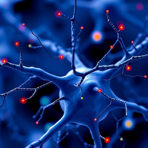
Ephaptic coupling, a form of neuron-to-neuron communication that operates without conventional synapses, is moving from the margins of neuroscience toward the center of a rapidly expanding research frontier. A new systematic review in BMC Bioinformatics examines nearly seventy mathematical models developed to explain how neurons can influence one another through changes in the extracellular electric field. The review, led by Pavel Y. Kondrakhin and colleagues at Sirius University of Science and Technology, brings together decades of theoretical work and shows how a mechanism first discussed in the 1940s is now being investigated with increasingly detailed computational tools. Its implications reach from ordinary brain rhythms and sensory processing to epilepsy, demyelination, neuropathic pain and neurodegenerative disease.
The word “ephaptic” refers to electrical interaction that occurs outside the specialized junctions formed by chemical or electrical synapses. When a neuron fires, currents flow along its membrane and through the surrounding extracellular space. Those currents alter the local extracellular potential, and nearby neurons can detect the resulting change because a neuron’s effective electrical state depends on the voltage difference between its interior and its surroundings. In simplified terms, if the extracellular voltage near a neighboring neuron shifts, the transmembrane voltage—the quantity that determines whether the cell moves toward or away from firing—also changes. The effect is usually subtle, but when many cells are densely packed, aligned in parallel or firing together, these small perturbations can accumulate and influence network behavior.
The mathematical models reviewed in the study span a wide range of complexity. At one end are phenomenological systems that reduce each neuron to a small number of variables, representing such features as membrane voltage, recovery processes and firing thresholds. These models are computationally efficient and can simulate large populations, making them useful for studying synchronization and collective rhythms. Quadratic integrate-and-fire models with ephaptic coupling, for example, describe neurons through simplified voltage dynamics while adding terms that represent the influence of an extracellular field. Although such models omit many biological details, they can reveal how weak electrical interactions alter the timing of spikes across thousands or millions of cells.
At the other end are biophysically detailed models based on cable theory and conductance-based descriptions of neuronal membranes. These frameworks divide an axon or dendrite into spatial compartments and calculate how electrical signals move through each segment. They can incorporate voltage-gated sodium and potassium channels, membrane capacitance, intracellular resistance and the geometry of the extracellular environment. Some models distinguish between myelinated and unmyelinated fibers, account for axonal diameter and represent the irregular placement of nodes of Ranvier, the exposed membrane regions where action potentials are regenerated. By including these features, researchers can ask whether ephaptic effects are amplified at specific anatomical structures or under particular patterns of neuronal activity.
A central technical challenge is determining how the extracellular field should be calculated. In the simplest approaches, the field is approximated by assuming that neurons are embedded in a uniform conductive medium. More advanced simulations solve the electrical interactions between extracellular and intracellular spaces directly. Extracellular–membrane–intracellular, or EMI, models describe the geometry of cells and the surrounding medium together, allowing researchers to examine how currents are redistributed near membranes. Kirchhoff–Nernst–Planck models go further by representing ionic movement, electric forces and concentration gradients. These approaches are computationally demanding, but they may become essential when ephaptic coupling is linked to changes in extracellular sodium, potassium or other ion concentrations.
The review highlights why ephaptic coupling is attracting attention in studies of synchronization. Conventional synaptic communication typically introduces delays, nonlinear chemical transformations or specific connection patterns. Ephaptic interactions, by contrast, can influence nearby cells almost instantaneously through the shared electric environment. A neuron’s extracellular field may slightly advance or delay the firing of another neuron, and repeated interactions can align spike timing. In a densely organized bundle of axons, this feedback may create a form of electrical cooperation: synchronized activity strengthens the local field, which in turn makes further synchronization more likely. The result could help explain some population-level rhythms observed in electroencephalography, electrocorticography and local field-potential recordings.
The same mechanism may also affect how nervous systems encode sensory information. Sensory pathways often contain tightly packed, similarly oriented fibers in which electrical fields can spread efficiently. A stimulus that activates many neighboring axons could therefore modify the timing and reliability of signals without changing the chemical synapses connecting those neurons. Small shifts in spike timing can carry important information, particularly in systems that use temporal coding. Mathematical models allow researchers to test whether ephaptic effects sharpen a population response, broaden it, create phase alignment or alter the threshold at which a stimulus becomes detectable. These predictions can then be compared with recordings from peripheral nerves, sensory circuits and cortical tissue.
Pathology provides another reason to take the phenomenon seriously. In epilepsy, large populations of neurons become excessively synchronized, producing abnormal electrical discharges that can spread through the brain. The reviewed models suggest that extracellular fields may participate in this process, either by reinforcing synchronization or by changing the conditions under which a seizure propagates. Demyelination could have especially important consequences because myelin normally insulates axons and shapes the distribution of current. When that insulation is damaged, current may leak into the extracellular space, potentially increasing unintended interactions between neighboring fibers. Such effects could contribute to conduction abnormalities in disorders such as multiple sclerosis, although the precise role of ephaptic coupling remains an active research question rather than a settled clinical explanation.
The review also connects ephaptic modeling with questions about neuropathic pain and neurodegeneration. Damage to axons, changes in membrane structure and altered ion concentrations can all modify the electrical environment around neurons. If injured fibers become more excitable or electrically exposed, neighboring fibers might be recruited in abnormal ways, potentially contributing to spontaneous activity or distorted sensory signals. In neurodegenerative conditions, the loss of tissue organization and changes in extracellular space may likewise reshape field-mediated interactions. The models do not establish that ephaptic coupling is the primary cause of these diseases, but they offer a way to investigate mechanisms that conventional synaptic diagrams may overlook.
By cataloguing the available models, their assumptions, tested hypotheses and open-source implementations, Kondrakhin and his colleagues aim to make the field easier to navigate. The review emphasizes that no single model can capture every level of ephaptic biology. Simplified equations are indispensable for exploring large networks and identifying general principles, while detailed simulations are needed to test how morphology, ion dynamics and tissue geometry shape the effect in real neurons. The next phase of research will likely depend on linking these approaches with experimental measurements of extracellular potentials, axonal structure and ionic composition. If that integration succeeds, ephaptic coupling could become more than a specialized theoretical concept: it could emerge as a measurable component of how neural circuits synchronize, compute and fail.
Cite Scienmag News
Cassandra Pierce. (August 25, 2026). Scientists Review Mathematical Models of Ephaptic Coupling Between Neurons. Scienmag. https://scienmag.com/scientists-review-mathematical-models-of-ephaptic-coupling-between-neurons/
Cassandra Pierce. "Scientists Review Mathematical Models of Ephaptic Coupling Between Neurons." Scienmag, 25 August 2026, https://scienmag.com/scientists-review-mathematical-models-of-ephaptic-coupling-between-neurons/. Accessed 4 September 2026.
Cassandra Pierce. "Scientists Review Mathematical Models of Ephaptic Coupling Between Neurons." Scienmag. August 25, 2026. https://scienmag.com/scientists-review-mathematical-models-of-ephaptic-coupling-between-neurons/


