In a groundbreaking study published in the prestigious journal npj Parkinson’s Disease, researchers have unveiled compelling evidence indicating that striatal hyperechogenicity observed through ultrasound imaging may serve as a critical biomarker for the prodromal phase of X-linked dystonia-parkinsonism (XDP). This discovery not only offers unprecedented insight into the pathophysiological underpinnings of XDP but also holds transformative potential for the early diagnosis and management of this debilitating neurodegenerative disorder. The study, led by Pauly et al., signifies a pivotal advance in the ongoing quest to decode the enigmatic progression of XDP through non-invasive imaging modalities.
X-linked dystonia-parkinsonism is a hereditary movement disorder predominantly affecting males, especially those of Filipino descent, characterized by the coexistence of dystonia and parkinsonism symptoms. Traditionally, diagnosis has been reliant on clinical criteria and genetic testing, which often recognize the disease only after the onset of overt motor symptoms. This lag in diagnosis severely limits timely therapeutic intervention. By focusing on striatal hyperechogenicity—an increased echogenic signal in the striatum region detected via transcranial ultrasound—the team has illuminated a novel, early biomarker that could detect the disease during its prodromal, or preclinical, phase.
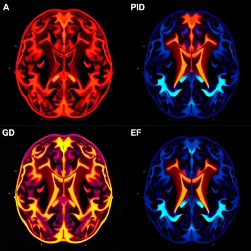
The study utilized advanced high-resolution transcranial ultrasound techniques to examine the brains of subjects genetically predisposed to XDP but not yet manifesting clinical symptoms. The striatum, a deep brain structure integral to motor control and implicated heavily in movement disorders, showed significant hyperechogenic signals in these prodromal individuals compared to matched controls. This finding suggests that pathological alterations in striatal tissue architecture and composition precede the clinical onset of XDP, providing a window of opportunity for early detection.
Crucially, the research delineates the pathological hallmarks that might contribute to hyperechogenicity in the striatum. The authors posit that iron accumulation within the striatal neurons and surrounding glial cells may alter the acoustic properties of the tissue, creating the hyperechogenic signature observed. This hypothesis aligns with existing studies linking abnormal iron metabolism to various neurodegenerative conditions, including Parkinson’s disease, underscoring a shared pathological pathway that might be exploited for diagnostic purposes across multiple disorders.
Beyond iron deposition, microstructural changes such as gliosis, neuronal loss, and inflammatory processes likely amplify the echogenic signal. The striatum’s vulnerability in XDP, exacerbated by genetic mutations on the X chromosome affecting the TAF1 gene, might trigger progressive neurodegeneration that subtly remodels the tissue even before clinically recognizable symptoms arise. These cumulative microscopic changes form the substrate for the ultrasound imaging markers identified by the study.
The implications of these findings extend far beyond early diagnosis. Striatal hyperechogenicity, as a measurable and quantifiable imaging marker, could allow clinicians to stratify at-risk populations, monitor disease progression objectively, and assess therapeutic responses in clinical trials. This is a critical breakthrough in a field where biomarkers have been sorely lacking, thus impeding efforts to develop targeted treatments and intervene before irreversible neuronal damage occurs.
Importantly, the utilization of transcranial ultrasound presents a highly accessible, cost-effective, and non-invasive diagnostic tool that could be adopted widely, particularly in resource-limited settings where XDP prevalence is highest. Compared to more complex neuroimaging modalities like PET or MRI, ultrasound offers a pragmatic solution suited for routine screening and longitudinal monitoring. This democratization of advanced diagnostic capability could fundamentally reshape the clinical landscape for XDP and similar disorders.
The study’s comprehensive approach involved a multidisciplinary team integrating expertise in neurology, radiology, genetics, and biomedical engineering. By correlating imaging findings with genetic and clinical data, the researchers constructed a robust framework linking striatal hyperechogenicity with the earliest stages of disease pathogenesis. This model provides a template for future investigations aiming to uncover prodromal markers in other neurogenetic disorders.
Furthermore, the temporal dynamics of striatal hyperechogenicity warrant further exploration. Whether this imaging signature remains stable, intensifies, or fluctuates throughout disease progression is a question of paramount clinical importance. Longitudinal studies tracking patients from prodromal to advanced stages will be indispensable in validating striatal hyperechogenicity as a reliable surrogate endpoint for disease activity and guiding optimal timing for intervention.
While the findings herald a promising avenue for early detection of XDP, several challenges remain. The specificity of striatal hyperechogenicity to XDP versus other conditions presenting with similar imaging patterns needs rigorous assessment. Overlapping echogenic profiles in disorders such as Parkinson’s disease and other movement syndromes may complicate differential diagnosis, necessitating the integration of imaging data with genetic and biomolecular markers to enhance diagnostic precision.
Ethical considerations also come to the fore when screening asymptomatic individuals for a progressive neurodegenerative disease. The psychological impact of identifying prodromal markers must be balanced against the current absence of definitive disease-modifying therapies. Nonetheless, as novel treatments emerge, early identification facilitated by imaging biomarkers like striatal hyperechogenicity will become increasingly critical.
In summary, the identification of striatal hyperechogenicity as an ultrasound marker for prodromal X-linked dystonia-parkinsonism represents a significant leap forward in the field of movement disorders. This research not only enriches our understanding of XDP pathophysiology but also confronts longstanding diagnostic challenges with an innovative, accessible imaging approach. Its implications resonate deeply within neurology and underscore the importance of multidisciplinary collaboration in tackling complex neurodegenerative diseases.
The study paves the way for a new era wherein neurodegenerative disorders may be detected, monitored, and ultimately treated earlier and more effectively than ever before. As the field advances, the integration of imaging biomarkers with genetic and clinical assessments promises to revolutionize patient care, transforming the prognosis for affected individuals and offering hope to at-risk families worldwide. The future of XDP diagnosis and management looks increasingly hopeful, propelled by the insights laid out in this pioneering research.
Ultimately, this investigation not only highlights a novel diagnostic avenue for XDP but also exemplifies the broader potential of ultrasound imaging in neurodegeneration. As research continues, it is conceivable that similar hyperechogenic markers could be discovered for other elusive neurological conditions, enhancing early detection strategies and opening new frontiers for therapeutic innovation. The convergence of cutting-edge imaging technology with genetic and molecular sciences heralds a transformative period for neurology, promising profound benefits for patients globally.
This landmark study by Pauly and colleagues sets a new standard in the use of non-invasive imaging to unravel the mysteries of neurodegenerative diseases and challenges researchers to further elucidate the complex interplay between genetic vulnerabilities and brain tissue alterations that underlie these devastating conditions.
Subject of Research: X-linked dystonia-parkinsonism and its prodromal biomarkers
Article Title: Striatal hyperechogenicity as an ultrasound imaging marker for prodromal X-linked dystonia-parkinsonism
Article References:
Pauly, M.G., Diesta, C.C.E., Cataniag, P. et al. Striatal hyperechogenicity as an ultrasound imaging marker for prodromal X-linked dystonia-parkinsonism. npj Parkinsons Dis. 12, 133 (2026). https://doi.org/10.1038/s41531-026-01418-4
Image Credits: AI Generated
