


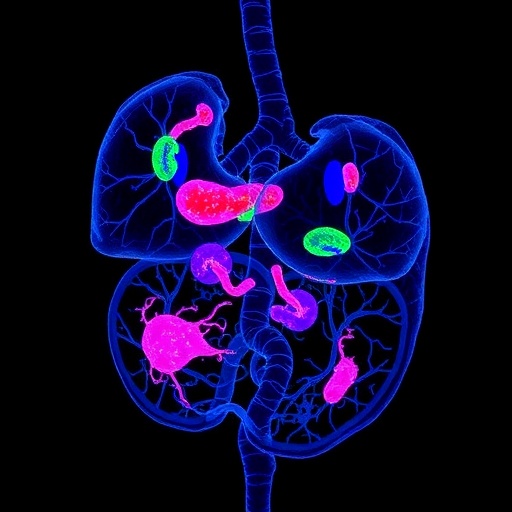
Researchers at Umeå University have unveiled groundbreaking insights into type 1 diabetes through a pioneering three-dimensional imaging study of an entire human pancreas. This research reveals that insulin-producing beta cells persist far longer after disease onset than had been previously recognized, fundamentally challenging the existing paradigm of beta-cell destruction in type 1 diabetes. The findings open new avenues for therapeutic intervention and deepen our understanding of the disease’s cellular complexity.
Utilizing state-of-the-art imaging technology, the team generated the first comprehensive 3D microscopic map of a whole pancreas obtained from a donor with late-onset type 1 diabetes. This achievement allowed them to visualize the intricate architecture of the pancreas in unprecedented detail. Contrary to longstanding beliefs that beta cells within the islets of Langerhans are completely destroyed, the study found a substantial presence of insulin-positive cells outside these traditional islet structures.
The researchers observed that beta cells were not only depleted within the pancreatic islets but also existed as isolated cells or small clusters distributed away from the main endocrine cell populations. This inverse spatial distribution—where extra-islet beta cells outnumber their islet-associated counterparts—is a revolutionary discovery, suggesting either an inherent resilience of these dispersed cells or the possibility of new beta-cell formation even in established diabetes.
By extending their gaze beyond the classical focus on islets, these scientists challenge the existing dogma and imply that conventional assessments may underestimate beta-cell survival. The beta-cell reservoir outside the islets offers a previously hidden dimension of cellular biology in diabetes, raising the hypothesis that the pancreatic microenvironment impacts beta-cell fate in complex and not yet fully understood ways.
Detailed three-dimensional imaging was key to achieving these insights. Traditional histological methods, which rely heavily on sectioned tissue samples, often miss dispersed cells located in less studied regions. The 3D approach enables the examination of individual cells throughout the entire organ, revealing spatial relationships and cellular configurations that were previously inaccessible. This holistic imaging strategy sets a new benchmark for pancreatic research and disease modeling.
The implications of this work extend beyond the basic science realm. The discovery of a significant extra-islet beta-cell population suggests novel targets for therapies aimed at preserving or even augmenting insulin-producing capacity. If these cells prove to be more resilient or capable of regeneration, harnessing their potential could revolutionize treatments, moving away from mere replacement therapies toward strategies that stabilize and empower the patient’s own residual beta cells.
Moreover, the researchers emphasize that the pancreatic microenvironment might hold clues to why some beta cells survive autoimmune attack. Understanding these protective niches could inform the design of microenvironment-targeted interventions, potentially halting or slowing the autoimmune progression hallmarking type 1 diabetes. This area of inquiry represents a frontier that may reshape therapeutic development strategies.
Joakim Lehrstrand, a doctoral candidate involved in the study, highlights that broadening the investigative focus beyond islets is essential for advancing beta-cell biology. This shift acknowledges the pancreas’s complex cellular landscape and underscores the importance of integrative approaches that combine imaging, molecular biology, and immunology to fully comprehend diabetes pathology.
The research group anticipates that whole-organ 3D imaging will become a core tool in future pancreas-related studies, including investigations into type 2 diabetes and pancreatic cancer. By enabling precise localization and isolation of distinct cell populations or regions, this method accelerates targeted molecular analyses and fosters a detailed understanding of pathological heterogeneity across diseases.
Ultimately, this study marks a milestone by demonstrating that beta-cell loss in type 1 diabetes is not as absolute as previously believed. The revelation of significant extra-islet beta-cell populations invites a reevaluation of disease timelines and treatment windows, suggesting new possibilities for intervention even after clinical diagnosis.
This exploration of pancreatic architecture and beta-cell distribution, published in Science Advances, heralds a paradigm shift with profound implications. It signals a move towards more nuanced views of pancreatic pathology and invites the scientific and medical communities to reconsider strategies for diagnosis, monitoring, and therapy in type 1 diabetes.
By integrating technological innovation with biological inquiry, the work from Umeå University exemplifies how precision mapping of organs can unravel hidden complexities in chronic diseases. Such advances promise to redefine our approach to some of the most challenging health issues facing humanity today.
News Publication Date: 22-May-2026
Web References: http://dx.doi.org/10.1126/sciadv.aed0496
Cite Scienmag News
Ophelia Keating. (May 26, 2026). 3D Whole-Organ Imaging Uncovers Residual Insulin-Producing Cells in Type 1 Diabetes. Scienmag. https://scienmag.com/3d-whole-organ-imaging-uncovers-residual-insulin-producing-cells-in-type-1-diabetes/
Ophelia Keating. "3D Whole-Organ Imaging Uncovers Residual Insulin-Producing Cells in Type 1 Diabetes." Scienmag, 26 May 2026, https://scienmag.com/3d-whole-organ-imaging-uncovers-residual-insulin-producing-cells-in-type-1-diabetes/. Accessed 3 September 2026.
Ophelia Keating. "3D Whole-Organ Imaging Uncovers Residual Insulin-Producing Cells in Type 1 Diabetes." Scienmag. May 26, 2026. https://scienmag.com/3d-whole-organ-imaging-uncovers-residual-insulin-producing-cells-in-type-1-diabetes/


