


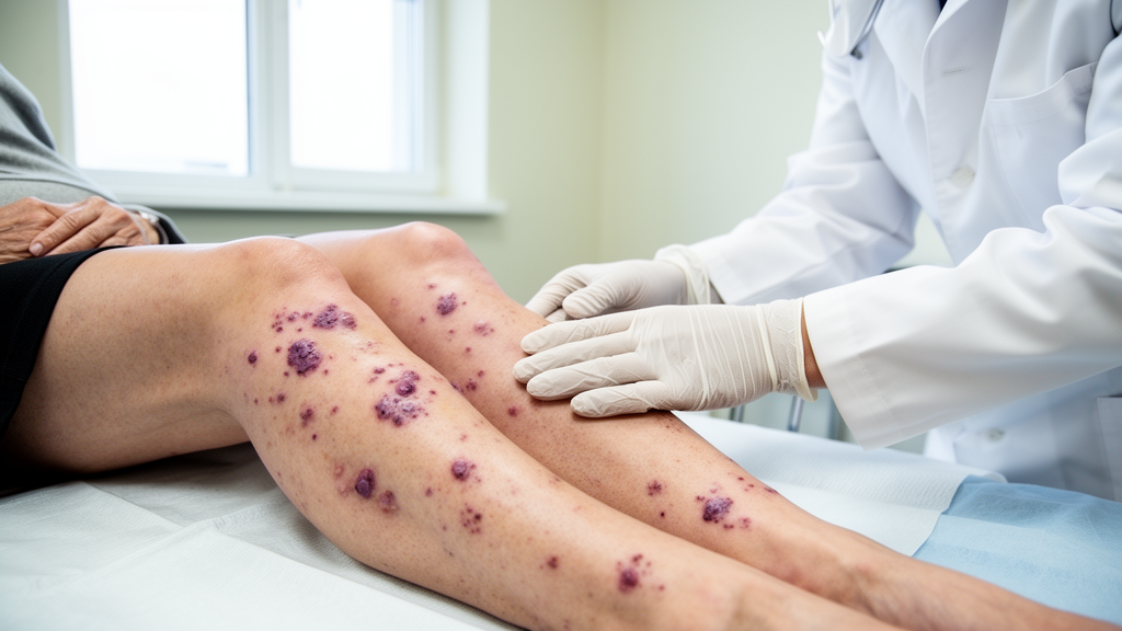
For dermatologists and rheumatologists, few diagnostic puzzles are as deceptively simple as palpable purpura. A patient presents with raised, reddish-purple lesions, a skin biopsy confirms leukocytoclastic vasculitis of the small vessels, and the workup begins. Yet the label cutaneous small-vessel vasculitis is only a starting point, because one of its most important underlying causes, immunoglobulin A vasculitis, carries very different implications for prognosis and follow-up. A new research letter published in the Archives of Dermatological Research by a team at The Ohio State University Wexner Medical Center suggests that clinicians may be able to separate these two entities earlier than previously thought, using nothing more exotic than the distribution of the rash, the patient’s age, and their smoking history.
Immunoglobulin A vasculitis, known for decades as Henoch-Schönlein purpura, is an immune-complex-mediated small-vessel vasculitis in which IgA-containing deposits accumulate in vessel walls, triggering inflammation and hemorrhage. It is the most common vasculitis of childhood, but it also occurs in adults, where its course tends to be more severe. The condition is defined by IgA1-dominant immune deposits detectable on direct immunofluorescence of skin or kidney biopsy tissue, and it classically involves not only the skin but also the joints, the gastrointestinal tract, and the kidneys. Renal involvement, which mirrors the pathology of IgA nephropathy, is the principal driver of long-term morbidity and is the reason clinicians monitor blood pressure, urinalysis, and estimated glomerular filtration rate in affected patients, sometimes for a year or more after the rash fades.
Generic cutaneous small-vessel vasculitis, by contrast, is a heterogeneous category. Its triggers include medications, infections, connective tissue diseases, and malignancies, and in many cases no cause is ever identified. When direct immunofluorescence shows no dominant IgA deposition, the prognosis is usually more benign, and the risk of glomerulonephritis is lower. The distinction therefore matters enormously: a patient with IgA vasculitis needs renal surveillance and rheumatology involvement, while a patient with drug-induced or idiopathic cutaneous vasculitis may need little more than removal of the offending agent and symptomatic care. The problem is that at the bedside, before biopsy results return, the two can look nearly identical.
The Ohio State team, led by dermatology principal investigator Abraham M. Korman with first author Hannah Moulton, approached the problem through a retrospective chart review approved by the university’s Institutional Review Board. The study compared adult patients diagnosed with IgA vasculitis against patients with cutaneous small-vessel vasculitis lacking IgA deposition, drawing on clinical data gathered across dermatology, rheumatology, and nephrology services. The collaboration reflects the multidisciplinary reality of vasculitis care, in which the dermatologist often makes the diagnosis, the rheumatologist manages systemic disease, and the nephrologist guards the kidneys. The work was supported in part by a Clinical and Translational Science Award from the National Center for Advancing Translational Sciences, and the authors report no competing interests.
The central finding is that three readily obtainable clinical variables, truncal lesion distribution, older age, and smoking status, help distinguish the two conditions. IgA vasculitis, in both its classic pediatric description and in adult series, has long been associated with lesions concentrated on the lower extremities and buttocks, gravity-dependent areas where hydrostatic pressure favors immune-complex deposition. Lesions extending onto the trunk point away from IgA vasculitis and toward other forms of small-vessel vasculitis. Conversely, the demographic and behavioral profile of the patients in each group differed in ways that clinicians can register within minutes of meeting the patient, long before laboratory results or immunofluorescence studies are available.
Age has been a recurring theme in the adult vasculitis literature. Prior studies, including a 2024 comparison of adult patients with cutaneous IgA vasculitis and non-IgA vasculitis published in Clinical and Experimental Dermatology by Gambichler and colleagues, and European consensus recommendations from the SHARE initiative published in Rheumatology in 2019, have emphasized that adult-onset IgA vasculitis behaves differently from the childhood disease, with higher rates of renal and gastrointestinal involvement and a more protracted course. The new research letter adds to this picture by suggesting that age itself, as a simple demographic variable, carries discriminative weight when clinicians are weighing which diagnosis best fits a given presentation of small-vessel vasculitis.
The association with smoking is perhaps the most intriguing of the three findings. Cigarette smoking has well-documented effects on the vasculature and on immune regulation, and it has been implicated in the expression of several inflammatory and autoimmune diseases. Its emergence as a distinguishing feature between IgA vasculitis and other cutaneous small-vessel vasculitides raises mechanistic questions that the research letter, by its nature as a brief report, does not fully resolve. Smoking could plausibly modify disease susceptibility, alter the phenotype of vasculitis once it develops, or simply correlate with other unmeasured exposures. Disentangling these possibilities will require larger, prospective cohorts, but the finding is a reminder that behavioral factors deserve a place alongside serology and histopathology in vasculitis research.
The practical implications extend to the diagnostic pathway itself. Direct immunofluorescence remains the gold standard for confirming IgA deposition, but biopsy and immunofluorescence take time, and the decision about how aggressively to pursue renal evaluation often must be made before those results return. European consensus recommendations already advise urinalysis and blood pressure monitoring at presentation and at regular intervals thereafter in suspected IgA vasculitis, and prior work by Hočevar and colleagues, published in Arthritis Research and Therapy in 2019, has sought to build predictive models for gastrointestinal and renal involvement in adults. Clinical variables that stratify risk at the first encounter, such as those highlighted in the new study, could help clinicians decide which patients warrant urgent nephrology input and closer laboratory surveillance while biopsy results are pending.
As with any retrospective, single-center study, the findings come with caveats. Chart reviews are vulnerable to selection bias and to variability in how diagnoses were coded and confirmed, and the data underlying the study, which contain protected health information, are not publicly available, though de-identified data may be shared upon reasonable request with approval from The Ohio State University Institutional Review Board. The results will need replication in larger and more diverse populations before they can be folded confidently into diagnostic algorithms. Even so, the message for practicing clinicians is concrete and immediately usable: when an adult presents with palpable purpura, the location of the lesions on the body, the patient’s age, and a careful smoking history are not incidental details of the social history. They are diagnostic signals, available at the bedside, that can begin to separate IgA vasculitis from the broader field of cutaneous small-vessel vasculitis and direct the workup toward the organs that matter most.
Subject of Research: Clinical differentiation of IgA vasculitis from cutaneous small-vessel vasculitis in adults
Article Title: Truncal lesions, age, and smoking distinguish IgA vasculitis from cutaneous small-vessel vasculitis
Article References: Truncal lesions, age, and smoking distinguish IgA vasculitis from cutaneous small-vessel vasculitis. (n.d.). https://doi.org/10.1007/s00403-026-04898-9
Image Credits: AI Generated
DOI: 10.1007/s00403-026-04898-9
Keywords: IgA vasculitis, cutaneous small-vessel vasculitis, leukocytoclastic vasculitis, direct immunofluorescence, palpable purpura, Henoch-Schönlein purpura, renal involvement, smoking, retrospective chart review, dermatology, rheumatology, diagnosis
Cite Scienmag News
Ophelia Keating. (October 9, 2026). Where the Rash Appears May Reveal Which Vasculitis a Patient Has. Scienmag. https://scienmag.com/where-the-rash-appears-may-reveal-which-vasculitis-a-patient-has/
Ophelia Keating. "Where the Rash Appears May Reveal Which Vasculitis a Patient Has." Scienmag, 9 October 2026, https://scienmag.com/where-the-rash-appears-may-reveal-which-vasculitis-a-patient-has/. Accessed 9 October 2026.
Ophelia Keating. "Where the Rash Appears May Reveal Which Vasculitis a Patient Has." Scienmag. October 9, 2026. https://scienmag.com/where-the-rash-appears-may-reveal-which-vasculitis-a-patient-has/


