


A 45-year-old man arrived at an emergency department in the United States with five days of worsening, 10-out-of-10 pain in his ankles, back, and shoulders. His home regimen of hydrocodone-acetaminophen no longer controlled the agony, which he recognized as the signature of a sickle cell pain crisis. Laboratory evaluation revealed severe normocytic anemia with a hemoglobin of 5.4 g/dL, far below a level any clinician would consider safe. Normally, the answer would be simple: transfuse packed red blood cells. But for this patient, the standard answer was unavailable. Two months earlier, following a hospitalization during which he received three crossmatched units of blood, his immune system had mounted a rare and complex alloantibody response that rendered virtually all compatible blood products inaccessible. The case, published in Frontiers in Hematology by a team from Western Michigan University Homer Stryker MD School of Medicine, illustrates an underrecognized clinical scenario in which the two pillars of sickle cell anemia care, transfusion and hydroxyurea, were simultaneously out of reach.
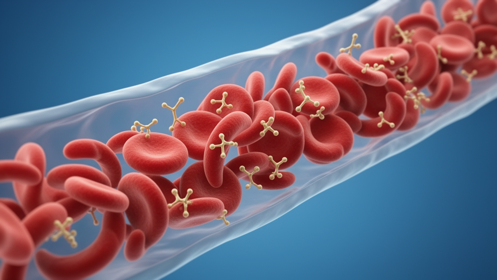
Sickle cell anemia is an autosomal recessive disorder caused by a single mutation in the β-globin gene that replaces normal hemoglobin A with abnormal hemoglobin S. When deoxygenated, hemoglobin S polymerizes into long chains that distort red blood cells into their characteristic sickle shape. These rigid, misshapen cells lodge in capillaries, producing the excruciating vaso-occlusive crises that can culminate in stroke, avascular necrosis, nephropathy, retinopathy, and venous thromboembolism. Because the disease also destroys red blood cells faster than the bone marrow can replace them, patients frequently present with acute anemia requiring transfusion. Yet transfusion is a double-edged sword. According to the literature cited in the report, between 15 and 50 percent of sickle cell patients develop alloantibodies against donor red cell antigens, antibodies that can trigger delayed hemolytic transfusion reactions and make every subsequent unit of blood harder to match. Complex alloimmunization can even preclude patients from curative therapies such as hematopoietic stem cell transplantation and gene therapy, which depend on safe transfusion support.
The story of this patient’s immunologic transformation began two months before his emergency presentation. Hospitalized for an acute sickle cell crisis with symptomatic anemia at a hemoglobin of 7.1 g/dL, he received three crossmatched units of packed red blood cells. Shortly after transfusion, he developed a febrile non-hemolytic transfusion reaction, a two-degree temperature spike with tachypnea requiring rapid response. At first, no overt hemolysis was evident. But over the following days, his hemoglobin fell progressively to 4.7 g/dL, dropping below his pre-transfusion baseline and raising concern for hyperhemolysis syndrome or warm autoimmune hemolytic anemia. The laboratory picture was dramatic: lactate dehydrogenase, a marker of cell destruction, surged from 529 U/L before transfusion to 1895 U/L afterward, while hemoglobin electrophoresis showed donor hemoglobin A declining from 34 percent to 19 percent as hemoglobin S rose from 59 to 73 percent, evidence that both transfused and native red blood cells were being destroyed. Reticulocyte counts remained elevated, indicating the marrow was working furiously to compensate.
Serologic detective work followed. The patient had previously undergone red blood cell genotyping demonstrating antibodies against D, E, C, and Kell antigens, a relatively common pattern in heavily transfused sickle cell patients. But subsequent evaluation at an outside reference laboratory revealed broad reactivity without an identifiable specificity, and eventually a newly formed anti-Fy3 alloantibody emerged. Extended genotyping delivered the crucial finding: the patient was homozygous for the FY*02N.01 (c.-67T>C) GATA-associated Duffy-null promoter variant, predicting an Fy(a-b-) erythrocyte phenotype. This promoter mutation disrupts GATA-1-mediated erythroid expression of the ACKR1 gene, abolishing Duffy antigen display on red blood cells. Herein lies the immunohematologic subtlety that makes this case remarkable. The Duffy-null phenotype is erythroid-restricted, not a complete absence of Duffy expression. The Duffy antigen/receptor for chemokines remains expressed in non-erythroid tissues, including endothelial cells. Consequently, individuals with this genotype are generally not expected to form anti-Fyb despite serologically typing Fy(b-), because Fyb is expressed outside erythrocytes. Anti-Fy3, directed against a common epitope on the FY protein separate from the Fya/Fyb polymorphism, is a different and clinically significant phenomenon, and patients carrying the GATA promoter mutation remain at risk for it.
How rare is anti-Fy3 in this setting? The Duffy-null phenotype itself is anything but rare. Howes and colleagues showed that the silent FYBES variant commonly reaches fixation across stretches of sub-Saharan Africa, and the phenotype is highly prevalent in populations of African ancestry. Yet Stone and colleagues noted only five prior case reports of acute or delayed hemolytic transfusion reactions due to anti-Fy3, and more recently Lemaire and Waldvogel Abramowski described three additional sickle cell disease patients who developed anti-Fy3 alloimmunization despite being homozygous for FY02N.01, two of them described as being in a transfusion deadlock. The paradox is striking: a genotype shared by millions becomes a transfusion trap in only a handful of documented patients. When this patient’s sample demonstrated additional unresolved reactivity, further hematologic evaluation ultimately identified anti-Jsa and anti-Doa alongside anti-Fy3, and compatible blood products became entirely unavailable.
The donor supply problem explains why. Anti-Fy3 requires Fy(a-b-) units for transfusion, and such units are found almost exclusively among donors of African ancestry. Hirani and colleagues reported Fy(a-b-) in just 0.2 percent of donors with Fy antigen testing in their regional analysis, rising to 2.6 percent among tested donors born in Africa. Layer on anti-Jsa, directed against a low-prevalence Kell-system antigen, and anti-Doa, directed against an antigen so uncommon that few donor units are even screened for it, and the compatibility search collapses. Anti-Doa carries a particular warning from the literature: it has been associated with recurrent hemolytic transfusion reactions not detected by standard crossmatching, meaning even serologically compatible units could prove dangerous. Hematology therefore recommended that future transfusions for this patient be reserved for life-threatening emergencies and require both cytopathologist and blood bank approval.
Facing a patient in severe crisis with a hemoglobin of 5.4 g/dL, an appropriate reticulocytosis of 17.0 percent, significant sickling on peripheral smear, and mild hemolysis markers, the clinical team pivoted to a transfusion-free strategy grounded in the American Society of Hematology 2020 guidelines. Those guidelines suggest immunosuppressive therapy with intravenous immunoglobulin, corticosteroids, and/or rituximab for sickle cell patients who have an acute need for transfusion but for whom compatible blood cannot be identified. The team administered a single 400 mg/kg dose of intravenous immunoglobulin, intravenous methylprednisolone at 40 mg every 12 hours transitioned to an oral prednisone taper, and two doses of subcutaneous epoetin alfa at 10,000 units to stimulate endogenous red cell production. Pain was managed with hydromorphone patient-controlled analgesia alongside continuous intravenous fluids, with folic acid, vitamin B6, vitamin B12, and vitamin C supplementation added per hematology recommendations. Hydroxyurea, the standard disease-modifying agent, was not pursued because of a history of myelosuppression, compounding the therapeutic inaccessibility.
The pharmacologic logic of this combination is elegant. Intravenous immunoglobulin likely works by blocking Fcγ receptors on macrophages, reducing the removal of antibody-coated red blood cells from circulation. It may also saturate the neonatal Fc receptor, FcRn, increasing clearance of circulating alloantibodies, and neutralize them through anti-idiotypic interactions. Corticosteroids inhibit Fc receptor-mediated clearance of sensitized red blood cells and, over time, dampen alloantibody production. Epoetin alfa, meanwhile, attacks the other side of the equation, driving the patient’s own marrow to replace red cells and reducing reliance on transfusion in someone for whom additional blood products carried high risk. Together, the three therapies targeted both ongoing immune-mediated destruction and insufficient red cell production. The team also considered mitapivat, a pyruvate kinase activator shown to reduce hemolysis by increasing ATP within red blood cells, but transfer to a tertiary care center for this experimental intervention was not possible due to limited bed availability.
The outcome was a quiet clinical success. During hospitalization, the patient’s hemoglobin stabilized between 6.0 and 6.5 g/dL and his pain gradually improved. On hospital day eight, he was transitioned from patient-controlled analgesia to oral oxycodone-acetaminophen with intravenous hydromorphone available for breakthrough pain. Although he remained fatigued from persistent anemia, he was clinically stable and felt prepared to manage his symptoms at home. He was discharged with a pain regimen and a plan for close outpatient follow-up, including weekly hemoglobin checks. The authors are candid about the limitations of their report: it describes a single patient, the relationship between the febrile non-hemolytic reaction and the identified alloantibodies was not definitively established, follow-up was limited, and long-term outcomes remain unavailable.
Beyond one patient, the case carries a broader message about transfusion stewardship. Current guidelines recommend prophylactic Rh and K antigen matching for all sickle cell patients, even in the absence of alloantibodies, to reduce the risk of sensitization. For patients who do develop alloantibodies, extended phenotype matching and molecular genotyping of donor units may prevent further alloimmunization, though the limited availability of such units must be weighed against the danger of delaying necessary transfusions. The authors call for further research into evidence-based management strategies for sickle cell patients with rare alloantibodies that preclude transfusion. As cell and gene therapies for sickle cell disease advance, safe transfusion support becomes more critical than ever, and cases like this one, where a common genotype produces an uncommon immunohematologic catastrophe, demand that clinicians recognize anti-Fy3 as a serious threat rather than a theoretical footnote.
Subject of Research: Hematology
Article Title: Case Report: Sickle cell anemia with complex alloimmunization
Article References: Zou, H., Alnounou, A., Kravchenko, T., Smeltzer, M., & Schaecher, C. (2026). Case Report: Sickle cell anemia with complex alloimmunization. Frontiers in Hematology, 5, Article 1870201. https://doi.org/10.3389/frhem.2026.1870201
Image Credits: AI Generated
DOI: 10.3389/frhem.2026.1870201
Keywords: Case, Report, Sickle, cell, anemia, complex, alloimmunization, scientific research
Cite Scienmag News
Drew Townsend. (October 17, 2026). When Compatible Blood Vanishes: The Rare Antibody Trap Inside Sickle Cell Anemia. Scienmag. https://scienmag.com/when-compatible-blood-vanishes-the-rare-antibody-trap-inside-sickle-cell-anemia/
Drew Townsend. "When Compatible Blood Vanishes: The Rare Antibody Trap Inside Sickle Cell Anemia." Scienmag, 17 October 2026, https://scienmag.com/when-compatible-blood-vanishes-the-rare-antibody-trap-inside-sickle-cell-anemia/. Accessed 17 October 2026.
Drew Townsend. "When Compatible Blood Vanishes: The Rare Antibody Trap Inside Sickle Cell Anemia." Scienmag. October 17, 2026. https://scienmag.com/when-compatible-blood-vanishes-the-rare-antibody-trap-inside-sickle-cell-anemia/


