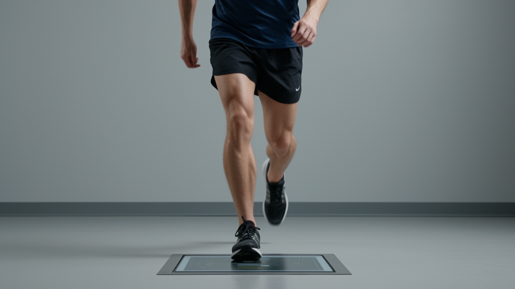
When healthy young adults sped up on a treadmill from a slow 0.5 metres per second to a moderate 1.0 metres per second, their muscles reorganised their activity in a widespread, distributed pattern, yet the ground reaction forces recorded beneath their feet showed no statistically significant waveform-level differences at all. That is the central finding of a new study published in BMC Biomedical Engineering, which set out to demonstrate an integrated analytical pipeline that combines statistical parametric mapping, functional principal component analysis, explainable machine learning, and equivalence testing within a single framework for multimodal gait signals.
The study, led by Kofi Nyantakyi Appiah with Edward Wilson Ansah and Frank Sarfo Dofour, analysed a publicly available multimodal gait dataset of healthy young adults. Fifty-eight participants contributed ground reaction force data, and fifty-five with complete electromyography recordings yielded 109 trials, 56 at the slow speed and 53 at the fast speed, for the electromyography and machine-learning analyses. The researchers drew on force-plate recordings from a split-belt treadmill sampled at 1000 hertz and surface electromyography from six lower-limb muscles: tibialis anterior, medial and lateral gastrocnemius, vastus lateralis, rectus femoris, and semitendinosus. Each stride was time-normalised to 101 points across the gait cycle, and forces were normalised to body mass.
The analytical core of the work was a paired statistical parametric mapping approach with sign-flip cluster-based permutation testing, using 5000 permutations to control the family-wise error rate. Rather than extracting discrete summary metrics such as peak values, this method treats the entire gait-cycle waveform as a continuous functional outcome, preserving temporal structure and allowing the detection of localised differences that discrete measures might miss. The authors also applied false discovery rate correction across the nine comparisons, six muscles and three force components, and the results were unchanged.
For the ground reaction forces, the outcome was a null result with an important nuance. No significant cluster-level differences were found for the vertical, anterior-posterior, or mediolateral force components. Conventional two one-sided tests applied to the vertical force waveform at each of the 101 time points, with equivalence bounds of plus or minus 0.2 newtons per kilogram, did not confirm practical equivalence either; mean differences ranged from minus 0.18 to plus 0.14 newtons per kilogram, and the null hypothesis of non-equivalence could not be rejected at any time point. The authors were careful to interpret this as an absence of evidence for a waveform difference, rather than evidence of preservation or equivalence. Notably, discrete outcomes such as the first vertical force peak did increase with speed, from 10.2 to 11.5 newtons per kilogram, confirming that the dataset was sensitive to speed and that the null mapping result reflected waveform-shape conservation rather than general insensitivity.
The electromyography results told a very different story. Significant clusters appeared in every muscle examined: ten clusters in tibialis anterior with effect sizes ranging from 0.44 to 0.61, along with clusters in both heads of the gastrocnemius, vastus lateralis, rectus femoris, and semitendinosus. The most pronounced speed-related increases occurred during early stance for tibialis anterior and vastus lateralis, mid-stance for the gastrocnemii, terminal stance and pre-swing for rectus femoris and semitendinosus, and swing for tibialis anterior. Several clusters were brief, spanning only two to four percent of the gait cycle, and the authors cautioned that cluster boundaries should be read as indicators of localised waveform divergence rather than precise physiological onset or offset times.
To translate these waveforms into a classification problem, the team used functional principal component analysis to compress each waveform into two principal component scores, yielding twelve features across the force axes and five muscle modalities. These features fed a Random Forest classifier with 1000 trees, evaluated with leave-one-subject-out cross-validation in which the eigenfunctions were computed strictly within each training fold to prevent data leakage. The classifier distinguished slow from fast walking with 87.2 percent accuracy, a 95 percent confidence interval of 79.3 to 92.3 percent, sensitivity of 90.6 percent, specificity of 83.9 percent, and an area under the curve of 0.92. A label-permutation test with 1000 permutations confirmed the accuracy significantly exceeded chance, and the result improved on simple amplitude features by 14.7 percentage points, with those features reaching only 72.5 percent accuracy.
Explainability came through SHAP, or SHapley Additive exPlanations, which quantified each feature’s contribution to the model’s decisions. Tibialis anterior principal component one ranked as the most influential feature, followed by pooled gastrocnemius and semitendinosus components. The loading curve for the tibialis anterior component captured overall amplitude modulation across the gait cycle, with positive loadings during swing phase, suggesting that the primary discriminative signal was the magnitude of dorsiflexion activity during swing, consistent with the foot-clearance demands of faster walking. The authors stressed that these SHAP values came from a post-hoc model trained on whole-cohort scores and are exploratory, to be interpreted alongside rather than instead of the waveform-level statistical findings.
The study’s scope and limitations deserve clear statement. The sample comprised healthy young adults with a mean age of 24 years, and the authors explicitly frame the work as a reference baseline and methodological demonstration; direct extrapolation to clinical populations such as stroke or Parkinson’s disease requires future validation. Electromyography amplitudes were not normalised to maximum voluntary contractions because the public dataset lacked reference recordings, which precludes comparing absolute activation levels across muscles and means the interpretations are limited to within-muscle relative changes and waveform timing. The analysis covered only the left leg, the speed range was narrow, and knee kinematics and EEG data from the original dataset were not used. Surface electromyography from tibialis anterior may also be contaminated by cross-talk from adjacent muscles.
Why does this matter beyond the treadmill laboratory? Gait speed is a key clinical indicator in neurological and orthopaedic conditions, and waveform-level adaptations in ground reaction forces and multi-muscle electromyography have remained poorly characterised because most prior work analyses discrete outcomes or individual modalities in isolation. By integrating continuous waveform inference, dimensionality reduction, explainable machine learning, and equivalence testing, the framework shows how statistical non-significance and practical equivalence can be distinguished, how latent temporal structure can be quantified, and how feature importance can be made transparent rather than hidden inside a black-box model. The authors note the offline pipeline takes roughly five minutes per participant on a standard desktop, feasible for research but requiring optimisation for real-time applications such as wearable biofeedback.
The authors position the pipeline as a foundation for future investigations in rehabilitation engineering, digital biomarkers, and wearable sensing, while emphasising that clinical utility in patient cohorts, real-world walking conditions, and longitudinal outcome studies remains to be tested. They also suggest that future work should include a broader range of walking speeds, since more pronounced muscle activation effects emerge at higher speeds than the 0.5 to 1.0 metres per second range studied here. For now, the message from this methodological demonstration is strikingly simple: when walking speed rises modestly, the nervous system redistributes muscle activity across many muscles and phases of the gait cycle, while the overall shape of the forces exchanged with the ground remains remarkably stable, at least within the sensitivity limits of this analysis.
Subject of Research: Multimodal analysis of ground reaction force and muscle activation waveforms during slow versus fast treadmill walking in healthy young adults
Article Title: Integrating statistical parametric mapping, functional principal component analysis, explainable machine learning, and equivalence testing for multimodal gait signal analysis
Article References: Appiah, K. N., Ansah, E. W., & Dofour, F. S. (2026). Integrating statistical parametric mapping, functional principal component analysis, explainable machine learning, and equivalence testing for multimodal gait signal analysis. BMC Biomedical Engineering, 8(1), Article 15. https://doi.org/10.1186/s42490-026-00118-7
Image Credits: AI Generated
DOI: 10.1186/s42490-026-00118-7
Keywords: gait analysis, electromyography, ground reaction force, statistical parametric mapping, machine learning, SHAP, functional principal component analysis, walking speed, treadmill, biomechanics, equivalence testing, digital biomarkers
Cite Scienmag News
Gregory Coleman. (October 28, 2026). Walking a little faster changes many muscles at once, but not the forces beneath your feet. Scienmag. https://scienmag.com/walking-a-little-faster-changes-many-muscles-at-once-but-not-the-forces-beneath-your-feet/
Gregory Coleman. "Walking a little faster changes many muscles at once, but not the forces beneath your feet." Scienmag, 28 October 2026, https://scienmag.com/walking-a-little-faster-changes-many-muscles-at-once-but-not-the-forces-beneath-your-feet/. Accessed 28 October 2026.
Gregory Coleman. "Walking a little faster changes many muscles at once, but not the forces beneath your feet." Scienmag. October 28, 2026. https://scienmag.com/walking-a-little-faster-changes-many-muscles-at-once-but-not-the-forces-beneath-your-feet/