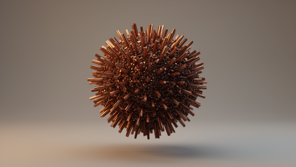
Materials chemists have grown microscopic particles that look like sea urchins and carry two different copper oxide crystal phases inside a single sphere. The new study, published in Applied Microscopy, describes urchin-like CuO/Cu2O mixed-phase microspheres roughly two to three micrometers across, each built from a dense core and radially aligned nanoneedles. By combining X-ray diffraction, X-ray photoelectron spectroscopy, high-resolution transmission electron microscopy, selected-area electron diffraction, and energy-dispersive spectroscopy, the team mapped where each phase resides and how the particles transform when bombarded by an electron beam.
The synthesis itself is deliberately simple. Two separate 5 millimolar aqueous solutions, one of copper (II) nitrate hydrate and one of hexamethylenetetramine, were sonicated for ten minutes, mixed in a 210 milliliter container, and treated with a dropwise-added surfactant blend of sodium hexadecyl sulfate, chloroform, and oleylamine. After thirty minutes of exposure to ambient air, the mixture was heated on a hot plate at 80 degrees Celsius for three hours and then cooled for ten minutes. The resulting nanostructures were transferred onto silicon wafers coated with silicon dioxide for analysis. According to the authors, the surfactant acts as a capping agent that steers anisotropic growth into radial nanoneedles while keeping the morphology uniform across the batch.
Scanning electron microscopy confirmed the urchin architecture at multiple magnifications. Low-magnification images showed large-scale uniformity, while higher magnifications revealed that each microsphere, averaging about two to three micrometers in diameter, consists of numerous radially aligned nanoneedles. That geometry creates a hierarchical architecture with a high surface-to-volume ratio, the property that makes such structures attractive for surface-driven applications. The authors note that conventional copper oxides in bulk or simple nanoparticle form often suffer from limited specific surface areas and rapid charge carrier recombination, limitations that hierarchical geometries and mixed-phase designs are meant to overcome.
X-ray photoelectron spectroscopy provided the first chemical evidence for phase coexistence. The Cu 2p spectrum showed the two main peaks for Cu 2p3/2 and Cu 2p1/2, along with strong satellite peaks at higher binding energies that are a characteristic fingerprint of Cu2+ in CuO. Deconvolution of the copper peaks and the O 1s spectrum, performed after Shirley background subtraction, confirmed the coexistence of Cu+ and Cu2+ species. The oxygen signals were assigned to Cu–OH at 531.1 electronvolts, CuO at 530.38 electronvolts, and Cu2O at 529.68 electronvolts. For these measurements, the urchin-shaped material was placed on a 50-nanometer-thick platinum-coated silicon substrate to minimize interference from the native oxide layer of the silicon.
X-ray diffraction then pinned down the crystal structures. The diffraction pattern showed distinct peaks indexable to both monoclinic CuO, the mineral tenorite, and cubic Cu2O, the mineral cuprite, assigned to the (110), (−111), (111), (202), (020), and (113) planes. The relatively broad peaks point to the nanocrystalline nature of the needle building blocks, and no impurity peaks were detected, which the authors interpret as confirmation of a high-purity mixed phase. Raman spectroscopy added a caveat: peaks at 295.75, 341.24, and 626.6 per centimeter matched monoclinic CuO vibrational modes, with the first two assigned to the Ag and Bg modes, but a distinct Cu2O Raman signature was not clearly resolved, possibly because of weak intensity, peak broadening, or overlap with the dominant CuO modes. The Raman result therefore primarily supports the CuO component, while the Cu2O contribution rests on the complementary techniques.
Transmission electron microscopy resolved the internal architecture in real space. Low-magnification images visualized the densely packed central core with radially protruding nanoneedles and an overall diameter near two micrometers. Selected-area electron diffraction on the peripheral needles produced well-defined diffraction rings characteristic of a polycrystalline structure, indexable to CuO crystallographic planes. The pattern from the central core instead showed a more discrete spot-like arrangement superimposed on the rings, suggesting higher crystallinity or aggregated nanocrystalline domains, a difference the authors attribute to the longer effective reaction time the core experiences early in the surfactant-assisted process. High-resolution TEM of a single nanoneedle revealed lattice fringes with an interplanar spacing of about 0.24 nanometers, matching the (111) plane of cubic Cu2O, and the continuity of those fringes over large regions indicates high crystallinity with minimal dislocations or stacking faults despite the rapid growth.
Atomic-resolution imaging reinforced that picture. High-angle annular dark-field scanning transmission electron microscopy, which differentiates regions by atomic density through Z-contrast, showed a periodic arrangement of copper atoms and confirmed a well-defined lattice in the copper oxide system. The absence of a significant amorphous layer on the needle surface suggests the surfactant-assisted synthesis stabilizes crystalline facets without leaving excessive organic residue behind.
The most striking result came from elemental mapping. Energy-dispersive X-ray spectroscopy on the HAADF-STEM image showed copper and oxygen distributed throughout the entire architecture, but their relative proportions varied systematically from center to rim. At the center, copper reached 79.72 atomic percent with oxygen at only 20.28 atomic percent, a copper-rich composition consistent with Cu2O or metallic copper clusters inferred from other analyses. At the edge, oxygen rose to 45.07 atomic percent while copper fell to 54.93 atomic percent, a ratio much closer to the stoichiometry of CuO, indicating that the radially grown nanoneedles are more highly oxidized. The team reads this spatial gradient as evidence for a core-first, oxidation-later mechanism: copper-rich seeds form the dense core first, and as the needles elongate under the surfactant template effect, their growing surface area allows more extensive oxidation through contact with the aqueous HMTA environment. The result is a spatially controlled heterostructure with a Cu2O-rich core and a CuO-rich shell and needle layer, a configuration the authors suggest could help create a built-in potential that assists charge carrier migration.
The particles also proved dynamic under the electron beam. In situ TEM at 200 kilovolts under continuous irradiation, with images acquired roughly every ten seconds, showed the urchin morphology gradually collapsing: the outer nanoneedles retracted and merged toward the central core until the hierarchical architecture gave way to a dense, compact particle. Because the beam current, absolute electron dose, and dose rate were not calibrated, the authors interpret the sequence qualitatively rather than as a quantitative dose-dependent transformation. They attribute the evolution to the high surface energy of the needle morphology, which lets surface atoms migrate under irradiation, driving surface reconstruction and a reduction in total surface energy. Inelastic electron scattering may also induce localized heating and radiolytic effects, potentially causing oxygen loss or partial phase transformation, while the observed volume shrinkage indicates that internal porosity from synthesis is eliminated through densification. In practical terms, the hierarchical architecture is metastable under high-energy irradiation.
Optical measurements rounded out the characterization. UV–visible absorption showed broad absorption extending through the visible region, characteristic of copper oxide and arising from the coexistence of the two phases with different band gaps: Cu2O typically around 2.0 to 2.2 electronvolts and CuO around 1.2 to 1.5 electronvolts. Taken together, the authors conclude that the combined XRD, XPS, SAED, EDS, and Raman evidence collectively supports mixed CuO/Cu2O phases within the microspheres, with strong visible-light absorption and a metastable but well-defined hierarchical structure. For a material built from earth-abundant, low-cost elements, that combination of phase control, spatial compositional gradient, and high surface area offers a structural framework for designing future functional materials, even as the electron-beam experiments caution that the delicate urchin geometry must be handled with its stability limits in mind.
Subject of Research: Mixed-phase structural characterization of urchin-like CuO/Cu2O nanostructures
Article Title: Mixed-phase structural characterization of urchin-like CuO/Cu₂O nanostructures
Article References: Jang, H. S., & Ryu, G. H. (2026). Mixed-phase structural characterization of urchin-like CuO/Cu₂O nanostructures. Applied Microscopy, 56(1), Article 19. https://doi.org/10.1186/s42649-026-00140-y
Image Credits: AI Generated
DOI: 10.1186/s42649-026-00140-y
Keywords: copper oxide, CuO, Cu2O, nanostructures, X-ray diffraction, transmission electron microscopy, X-ray photoelectron spectroscopy, energy-dispersive spectroscopy, electron-beam irradiation, hierarchical microspheres, surfactant-assisted synthesis, mixed-phase materials
Cite Scienmag News
Denise Maddox. (October 28, 2026). Urchin-Shaped Copper Oxide Microspheres Blend Two Crystal Phases in One Particle. Scienmag. https://scienmag.com/urchin-shaped-copper-oxide-microspheres-blend-two-crystal-phases-in-one-particle/
Denise Maddox. "Urchin-Shaped Copper Oxide Microspheres Blend Two Crystal Phases in One Particle." Scienmag, 28 October 2026, https://scienmag.com/urchin-shaped-copper-oxide-microspheres-blend-two-crystal-phases-in-one-particle/. Accessed 28 October 2026.
Denise Maddox. "Urchin-Shaped Copper Oxide Microspheres Blend Two Crystal Phases in One Particle." Scienmag. October 28, 2026. https://scienmag.com/urchin-shaped-copper-oxide-microspheres-blend-two-crystal-phases-in-one-particle/