


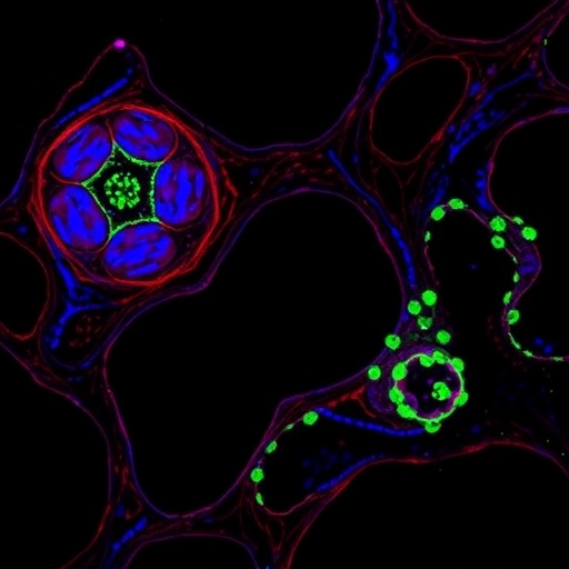
Liver cancer may be exploiting a molecular “sugar coating” to build the blood-vessel network it needs to grow, according to a study that identifies a previously underexplored connection between protein modification, cancer signaling and angiogenesis. Researchers report that the enzyme UGGT1 stabilizes the protein LGALS3BP through a process known as N-glycosylation, enabling tumor cells to activate the NOTCH signaling pathway and increase production of the vessel-promoting factor VEGF-A. In experiments using liver cancer cells, endothelial cells and mice, disrupting UGGT1 reduced tumor growth and blood-vessel formation. The findings suggest that the UGGT1–LGALS3BP pathway could become a target for anti-angiogenic therapies, although the work remains preclinical and has not yet demonstrated that blocking the mechanism is safe or effective in patients.
Liver cancer is among the world’s leading causes of cancer-related death, and hepatocellular carcinoma, or HCC, accounts for most primary liver cancers. One reason HCC is difficult to treat is its dependence on a highly adaptable tumor microenvironment. As tumors enlarge, they require oxygen and nutrients that cannot diffuse far through tissue. Cancer cells therefore release signals that encourage nearby blood vessels to sprout and connect with the tumor. This process, called angiogenesis, can also create disordered, leaky vessels that help malignant cells invade surrounding tissue and enter the circulation. Drugs that inhibit angiogenesis have improved outcomes for some patients, but tumors frequently develop resistance by activating alternative vascular signals. The new study, published in Clinical Proteomics, focuses on a molecular layer that may help explain how HCC sustains this vascular supply.
The central protein, LGALS3BP, is a secreted molecule that has been found at elevated levels in several malignant tumors, but its specific role in HCC angiogenesis has been unclear. The researchers first used bioinformatics analyses to examine LGALS3BP expression in liver cancer datasets and its relationship to angiogenesis-related genes and the NOTCH pathway. They then compared messenger RNA and protein levels in HCC tissues and cancer-cell models using quantitative reverse-transcription PCR and Western blotting. They also measured secreted LGALS3BP with enzyme-linked immunosorbent assays. Across these analyses, LGALS3BP was substantially more abundant in HCC tissue and cells than in corresponding noncancerous controls, supporting the idea that it is not merely a passive marker of malignancy but could be an active participant in tumor biology.
To test that possibility, the team reduced LGALS3BP production in HCC cells and measured several behaviors associated with aggressive disease. Loss of the protein diminished cell viability and proliferation, weakened the ability of cells to form colonies and reduced invasion through laboratory membranes in Transwell assays. The researchers also observed lower levels of VEGF-A, a powerful signal that binds receptors on endothelial cells and stimulates their migration, division and organization into new vascular structures. In a co-culture system, cancer cells were grown alongside human umbilical vein endothelial cells. When LGALS3BP was depleted, the endothelial cells formed fewer and less extensive tube-like networks, an in-vitro model of angiogenic activity. Together, the results linked LGALS3BP to both cancer-cell aggressiveness and the capacity of tumor cells to recruit vascular support.
The study then moved upstream to ask how LGALS3BP is regulated. The researchers identified an interaction between LGALS3BP and UGGT1, an enzyme located in the endoplasmic reticulum, the cellular compartment where many newly synthesized proteins are folded and processed. UGGT1, short for UDP-glucose:glycoprotein glucosyltransferase 1, functions as part of the endoplasmic-reticulum quality-control system. It recognizes incompletely folded glycoproteins and adds a glucose residue to specific N-linked carbohydrate chains, allowing the proteins to re-enter a cycle of binding to molecular chaperones. This quality-control process can give a protein additional opportunities to fold correctly before it is transported through the secretory pathway. Because LGALS3BP is secreted, its stability and maturation may be particularly sensitive to such processing.
Using protein-interaction analyses, co-immunoprecipitation and immunofluorescence microscopy, the researchers found evidence that UGGT1 associates with LGALS3BP. They next examined potential N-glycosylation sites—positions where a carbohydrate chain can be attached to an asparagine residue within a characteristic amino-acid sequence. LGALS3BP contains six candidate sites, and the team generated individual mutants in which the relevant asparagine was replaced, as well as a combined mutant lacking all six sites. These experiments showed that UGGT1-mediated glycosylation helped maintain LGALS3BP protein stability. When UGGT1 was reduced, LGALS3BP protein levels and secretion declined; restoring or increasing LGALS3BP could counter some of the downstream effects. The results point to a mechanism in which UGGT1 does not simply alter a signal at the cell surface, but helps preserve the supply of a secreted factor that cancer cells use to remodel their surroundings.
The downstream signaling route involved NOTCH, a communication system that helps neighboring cells coordinate decisions about development, survival and cell identity. NOTCH signaling begins when a ligand on one cell binds a NOTCH receptor on an adjacent cell. Proteolytic cleavage then releases the receptor’s intracellular domain, which travels to the nucleus and partners with DNA-binding proteins to switch target genes on or off. In blood-vessel formation, NOTCH helps regulate the balance between endothelial “tip” cells that lead a new sprout and “stalk” cells that follow and proliferate. Dysregulated NOTCH activity can therefore reshape the architecture and behavior of tumor-associated vessels. In the HCC models, LGALS3BP increased activity of NOTCH-associated molecules and raised VEGF-A expression, while suppressing LGALS3BP weakened this signaling axis. The findings support a UGGT1–LGALS3BP–NOTCH–VEGF-A chain connecting intracellular protein quality control to the formation of new tumor blood vessels.
The most demanding test came in animals bearing xenograft tumors, in which human HCC cells were implanted into mice. Reducing UGGT1 significantly slowed tumor growth and decreased markers of angiogenesis within the tumors. When LGALS3BP was overexpressed, some of the effects of UGGT1 loss were reversed, a result known as partial rescue. Such rescue experiments are important because they help place LGALS3BP downstream of UGGT1 rather than showing only that both proteins happen to be associated with aggressive tumors. Even so, partial rescue does not prove that the pathway is the only route by which UGGT1 influences cancer. UGGT1 participates in broad protein-folding quality control, and altering it could affect many glycoproteins at once. The apparent anti-tumor effect may therefore reflect a combination of LGALS3BP-dependent and independent changes.
The work raises the possibility of attacking liver cancer angiogenesis at a different point from existing VEGF-directed drugs. Directly blocking VEGF-A can starve tumors of vascular support, but cancer cells may compensate by increasing other growth factors or by changing the structure of their vessels. Inhibiting UGGT1 could, in principle, reduce the stability of several pro-tumor glycoproteins, while targeting LGALS3BP might offer a more focused way to interfere with the secreted signal identified in this study. Yet both strategies present challenges. UGGT1 is part of a normal cellular quality-control system, so systemic inhibition could harm healthy secretory tissues or trigger endoplasmic-reticulum stress. LGALS3BP is also expressed in noncancerous contexts, meaning that its role in immunity, tissue repair and other physiological processes will need to be defined before it can be safely targeted. The experiments used cell lines, endothelial co-cultures and xenograft models, which do not fully reproduce the immune system, genetic diversity or treatment history of human HCC.
The researchers describe their findings as evidence for a potential anti-angiogenic target, not as a ready-made therapy. The next steps will include confirming the pathway in larger collections of patient tumors, determining whether UGGT1 or LGALS3BP levels predict vascular activity or treatment response, and developing inhibitors that can distinguish cancer-relevant signaling from essential normal protein processing. It will also be important to test whether disrupting the pathway improves the performance of existing immunotherapies or molecular treatments, and whether tumors can bypass it through alternative angiogenic programs. For now, the study offers a striking mechanistic explanation for how a modification made inside the endoplasmic reticulum may influence events far outside the cancer cell: by stabilizing a glycosylated secreted protein, UGGT1 may help HCC turn on NOTCH and VEGF-A, recruiting the blood vessels that allow the disease to expand.
Cite Scienmag News
Nathaniel Bowman. (August 27, 2026). UGGT1 Drives Liver Cancer Angiogenesis by Glycosylating LGALS3BP Through NOTCH Signaling. Scienmag. https://scienmag.com/uggt1-drives-liver-cancer-angiogenesis-by-glycosylating-lgals3bp-through-notch-signaling/
Nathaniel Bowman. "UGGT1 Drives Liver Cancer Angiogenesis by Glycosylating LGALS3BP Through NOTCH Signaling." Scienmag, 27 August 2026, https://scienmag.com/uggt1-drives-liver-cancer-angiogenesis-by-glycosylating-lgals3bp-through-notch-signaling/. Accessed 3 September 2026.
Nathaniel Bowman. "UGGT1 Drives Liver Cancer Angiogenesis by Glycosylating LGALS3BP Through NOTCH Signaling." Scienmag. August 27, 2026. https://scienmag.com/uggt1-drives-liver-cancer-angiogenesis-by-glycosylating-lgals3bp-through-notch-signaling/


