


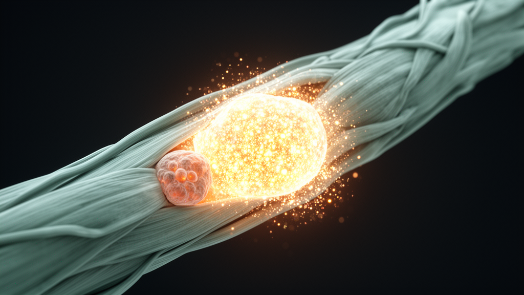
Skeletal muscle has a remarkable capacity for self-repair, but a new study suggests that one of the body’s most fundamental chemical messengers can quietly sabotage that process. Researchers report that when intracellular calcium levels remain chronically elevated, muscle progenitor cells get stuck in a proliferative state and never commit to becoming mature muscle fibers. The culprit, they show, is a sustained calcium-dependent signaling cascade centered on the transcription factor NFATc1, which keeps proliferation genes switched on at precisely the moment cells should be exiting the cell cycle and fusing into new myotubes. The findings, published in Experimental & Molecular Medicine, identify a molecular mechanism that may underlie muscle weakness in conditions as varied as aging, estrogen deficiency, and calcific tendinitis, and they point to NFAT inhibition as a potential therapeutic strategy.
The research began with a question that has lingered in the muscle biology literature for years. Calcium dysregulation is a well-documented feature of diseased and aging muscle. Patients with systemic hypercalcemia, defined by serum calcium exceeding 3.0 millimolar, frequently complain of fatigue and proximal muscle weakness. Duchenne muscular dystrophy drives chronic cytosolic calcium overload through loss of the structural protein dystrophin. Aging muscle, meanwhile, shows increased resting intracellular calcium, leaky ryanodine receptors, and inefficient calcium reuptake by SERCA pumps. Yet most research had focused on how calcium controls contraction in mature fibers, leaving the impact on regenerating muscle largely unexplored.
To connect those dots, the team, led by Kee K. Kim of Chungnam National University, first turned to human data. Analyzing transcriptomes from 679 individuals in the GTEx v8 dataset, they compared gastrocnemius muscle from people aged 50 and older with samples from those under 40. Gene set enrichment analysis revealed that aged muscle showed coordinated activation of calcium signaling pathways and muscle cell proliferation programs, alongside upregulation of the quiescence regulator SPRY1, the early myogenic factor PAX3, and the proliferation marker CCND1. Crucially, the signature appeared in both men and women, suggesting that a proliferative bias tied to calcium signaling is a general feature of aging muscle rather than a sex-specific phenomenon.
The researchers then moved into the laboratory to test whether calcium excess directly alters myoblast behavior. When C2C12 mouse myoblasts and primary myoblasts isolated from hindlimb muscle were exposed to elevated calcium chloride under normal growth conditions, nothing dramatic happened. But when the same cells were pushed to differentiate while bathed in 3 or 6 millimolar calcium chloride, a striking picture emerged: total cell numbers rose significantly, the proliferative marker PAX7 climbed at both the protein and mRNA levels, and the cells failed to exit the cell cycle. Calcium imaging with the Fluo-4 AM indicator confirmed that intracellular calcium remained persistently elevated throughout the differentiation window, rising in a dose-dependent manner in primary myoblasts.
That persistent proliferative state came at a direct cost to myogenesis. Myotube formation declined dose-dependently in calcium-exposed C2C12 cultures, and levels of the myogenic markers MYH3 and myogenin dropped alongside their messenger RNAs. Primary mouse myoblasts showed the same defects, and, importantly, the phenomenon crossed the species line: human embryonic stem cell-derived myoblasts differentiated for two weeks in 3 millimolar calcium chloride formed fewer myotubes and produced less total myosin heavy chain. Notably, prolonged exposure to lower concentrations sufficed to impair differentiation, indicating that the duration of calcium elevation, rather than its absolute peak, is the critical determinant of pathology.
RNA sequencing of calcium-exposed primary myoblasts identified 2,776 differentially expressed genes and revealed the underlying transcriptional logic. Genes upregulated by calcium exposure were enriched for cell division and chromosome segregation, while downregulated genes clustered around muscle contraction and sarcomere organization. Gene set enrichment analysis flagged activation of the calcium-dependent NFAT pathway. Mechanistic experiments followed: NFATc1, tagged with green fluorescent protein, translocated into the nucleus of calcium-exposed myoblasts; the NFAT target gene Ccnd1, encoding cyclin D1, was upregulated; and chromatin immunoprecipitation showed that NFATc1 binding to the Ccnd1 promoter was significantly enhanced under calcium excess. In short, calcium was hijacking a pathway that normally helps orchestrate differentiation and repurposing it to keep the cell-cycle engine running.
To confirm the mechanism in living animals, the team used two complementary models. The first was the ovariectomized mouse, in which estrogen deficiency destabilizes calcium handling by hyperphosphorylating the ryanodine receptor 1, provoking spontaneous calcium release from the sarcoplasmic reticulum. These mice lost bone density, performed worse on treadmill endurance tests, and developed gastrocnemius atrophy. Their muscle transcriptomes mirrored the in vitro signature: enrichment of calcium signaling and proliferation programs, downregulation of developing and mature myosin heavy chain genes, upregulation of Pax7 and MyoD, shrunken myofiber cross-sectional areas, and clear nuclear translocation of NFATc1. The second model was human calcific tendinitis. In supraspinatus muscle samples taken from patients during calcific deposit removal, tissue adjacent to the calcium deposits showed downregulation of SPRY1, PAX3, and MYH3 together with upregulation of CCND1 compared with remote regions, demonstrating that even localized calcium overload in human tissue produces the same proliferative, anti-myogenic fingerprint.
The most translational part of the study came from pharmacological rescue. Using VIVIT, a selective peptide inhibitor of NFAT, the researchers blocked calcium-induced nuclear accumulation of NFATc1 in both C2C12 cells and primary myoblasts. VIVIT restored MYH3 expression and reversed the myotube formation defect under calcium overload. A complementary experiment using caffeine to induce sarcoplasmic reticulum calcium leak recapitulated the suppression of myoblast fusion, and VIVIT rescued that too, confirming the effect arises from intracellular calcium dysregulation rather than nonspecific stress. In vivo, the team injured tibialis anterior muscles with barium chloride, which triggers calcium-dependent proteolysis and robust regeneration, and injected VIVIT locally on days 0 and 2. Treated muscles showed improved regenerative morphology, larger myofiber cross-sectional areas, and a dramatic reduction in the proportion of immature myogenin-positive fibers, all without measurable changes in inflammatory markers such as Il6, Tnfa, and Inos.
One conceptual takeaway is that NFAT plays a duration-dependent dual role in muscle. Transient calcium-dependent NFAT activation has classically been viewed as a positive regulator of myogenesis, supporting fiber-type specification and hypertrophy. The new data show that when calcium elevation persists, the same pathway flips into an anti-myogenic mode, sustaining cyclin D1 expression and preventing the cell-cycle exit that myoblasts must undergo to fuse into contractile fibers. The authors also suggest this framework may help explain osteosarcopenia, the poorly understood co-occurrence of osteoporosis and muscle loss, since both tissues depend on well-regulated calcium handling, although they caution that this connection remains inferential and requires direct testing.
Limitations remain, and the researchers are candid about them. The therapeutic benefit of VIVIT was demonstrated in an acute injury model, not in the chronic ovariectomy setting, because repeated intramuscular injections over a six-week protocol would introduce local tissue damage and inflammation that could confound the results. Future work with long-term pharmacological approaches or genetic models will be needed to validate NFAT as a drug target for chronic calcium-associated myopathies. Still, by tracing a single molecular thread from aged human muscle through mouse myoblasts, ovariectomized mice, calcific tendinitis patients, and a rescued injury model, the study establishes chronic calcium-NFAT signaling as a credible mechanism linking calcium dysregulation to failed muscle repair, and it offers a concrete starting point for therapies aimed at restoring the regenerative capacity of weakened muscle.
Subject of Research: Mechanism by which pathological calcium overload impairs skeletal muscle regeneration through sustained NFAT-dependent proliferative signaling
Article Title: Pathological calcium overload impairs skeletal muscle regeneration through sustained NFAT-dependent proliferative signaling
Article References: Lee, S., Cho, N., Jo, S., Ji, J.-H., Kim, J. O., Park, C., Choi, S., Kim, E.-M., & Kim, K. K. (2026). Pathological calcium overload impairs skeletal muscle regeneration through sustained NFAT-dependent proliferative signaling. Experimental & Molecular Medicine. https://doi.org/10.1038/s12276-026-01856-3
Image Credits: AI Generated
DOI: 10.1038/s12276-026-01856-3
Keywords: skeletal muscle, muscle regeneration, calcium signaling, NFATc1, myogenesis, satellite cells, cyclin D1, sarcopenia, calcific tendinitis, VIVIT, aging muscle, ovariectomy
Cite Scienmag News
Nathaniel Bowman. (October 9, 2026). Too Much Calcium Traps Muscle Stem Cells in Growth Mode, Blocking Repair. Scienmag. https://scienmag.com/too-much-calcium-traps-muscle-stem-cells-in-growth-mode-blocking-repair/
Nathaniel Bowman. "Too Much Calcium Traps Muscle Stem Cells in Growth Mode, Blocking Repair." Scienmag, 9 October 2026, https://scienmag.com/too-much-calcium-traps-muscle-stem-cells-in-growth-mode-blocking-repair/. Accessed 9 October 2026.
Nathaniel Bowman. "Too Much Calcium Traps Muscle Stem Cells in Growth Mode, Blocking Repair." Scienmag. October 9, 2026. https://scienmag.com/too-much-calcium-traps-muscle-stem-cells-in-growth-mode-blocking-repair/


