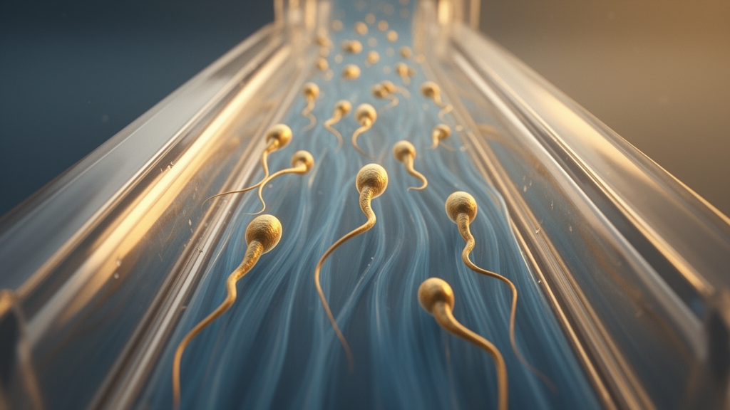
In a quiet revolution unfolding inside palm-sized chips of transparent polymer, scientists are learning to sort sperm the way the female body does: by letting the strongest swimmers fight the current. A new review published in Discover Fluid Mechanics by Dhiraj B. Puri, Harshit Wadhwa, Vadiraj Hemadri, Gautam Biswas and Siddhartha Tripathi surveys the physics of sperm rheotaxis — the tendency of sperm cells to orient and swim against fluid flow — and catalogs a growing family of microfluidic devices that exploit this behavior to isolate highly motile, viable sperm for assisted reproduction. The review, published on 24 February 2026, argues that rheotaxis offers a passive, non-invasive alternative to conventional sperm preparation methods that can damage the very cells clinics are trying to rescue.
The phenomenon itself is elegantly physical. Sperm are microscale swimmers operating at low Reynolds number, a regime where viscous forces utterly dominate inertia. Propulsion comes from a flexible flagellum whose internal axoneme — a characteristic 9 + 2 arrangement of microtubule doublets — is bent by ATP-driven dynein motor proteins into traveling waves. As these waves propagate from the midpiece toward the tail tip, anisotropic drag along the slender body produces a net thrust that pushes the cell forward. But swimming alone does not produce upstream navigation. That requires a second ingredient: a nearby surface. Sperm naturally accumulate at walls because of asymmetries in their beating — the flagellum swings with larger lateral amplitude than the head, generating a wall-directed lift force that keeps the cell parallel to the boundary.
Once a sperm is pinned near a wall and a shear flow is imposed, the head and tail occupy different heights within the velocity gradient and therefore experience unequal drag. This imbalance generates a hydrodynamic torque about the cell’s center of mass that rotates the sperm until it faces upstream. The angular dynamics can be captured by a simple torque-balance relation, dθ/dt = −Aγ sin θ, where θ is the angle between the sperm’s orientation and the flow direction, γ is the local shear rate, and A is a geometry-dependent constant. The reorienting torque vanishes when the cell is fully aligned or anti-aligned with the flow and peaks when it is perpendicular. Crucially, the mechanism is passive: classical hydrodynamic theory from Bretherton and Rothschild onward, and modern microfluidic experiments, converge on the view that sperm need not actively sense the flow at all.
The experimental record supports this passive interpretation in striking detail. Zhang and colleagues reported no detectable changes in flagellar waveform or intracellular calcium signaling during rheotactic alignment, suggesting sperm do not perceive flow direction. Yet cellular state still matters: Miki and colleagues showed that sperm lacking the CatSper calcium channel failed to maintain upstream trajectories, while Schiffer and colleagues found that passive biomechanical interactions can still enable rolling and rheotaxis even without CatSper-mediated calcium signaling. The chiral, handed nature of the flagellar beat adds another twist — Kantsler and colleagues demonstrated that sperm in controlled shear flows trace spiral or helical upstream trajectories, the result of a tug-of-war between shear-induced torque and the rotational bias of the beat itself.
Rheotaxis also operates only within a window. At very low shear, the reorienting torque is too weak to beat random rotational fluctuations; at excessively high shear, fluid forces simply sweep the cell downstream. Experiments place stable upstream motion for human sperm at flow velocities of roughly 22 to 102 micrometers per second, and for bovine sperm at 22 to 107 micrometers per second. El Sherry and colleagues found that 80 to 84 percent of sperm exhibited rheotaxis in a straight microfluidic channel across flow velocities of 33 to 134 micrometers per second, with reversal time inversely proportional to flow velocity. Fluid rheology matters too: Puri and colleagues showed that upstream progressive velocity decreased in Newtonian fluids but increased in non-Newtonian fluids as flow rate rose, and that increasing flow pushed sperm from the channel center toward the walls.
Why does this physics matter clinically? Infertility affects roughly one in six couples worldwide — about 17.5 percent of the adult population, according to the World Health Organization — and male factors contribute to about half of all cases. Assisted reproductive technologies such as in vitro fertilization, intrauterine insemination and intracytoplasmic sperm injection all depend on the availability of high-quality motile sperm. The two conventional preparation methods, density gradient centrifugation and the swim-up technique, bypass the natural selection barriers of the female reproductive tract and, studies indicate, can induce oxidative stress and DNA fragmentation in sperm, potentially impairing their fertilizing ability.
Rheotaxis-based microfluidics aims to restore that natural filtration. In one design strategy, geometric trapping and flow structuring do the sorting.
Zaferani and colleagues built a microchannel “corral” system with funnel-shaped barriers that force all cells through a narrow, high-shear zone; only rheotactic swimmers can then enter the low-flow collection region. The device contains seven interior corrals, ahead of each of which rheotactic zones are created by tuning the injection rates of the sperm medium, allowing motile cells to reorient, swim against the flow and become confined within the corral structure. Sarbandi and colleagues took a similar biomimetic approach, incorporating eight micro pockets on the channel walls that mimic the microgrooves of the female reproductive tract; the pockets progressively expand the streamlines, creating a sharp shear gradient in their frontal zone and a shear-protected region inside where viable, highly motile sperm accumulate. Both devices, the review notes, require dilution of the semen sample, since raw semen will clog the channels. Mane and colleagues proposed a simpler T-shaped design with one inlet and two outlets, one of which is sealed: the sealed channel produces a high velocity gradient and a rheotaxis zone at the junction, trapping separated sperm in low-Reynolds-number flow so they cannot re-enter the stream. The device separated more than 3,500 sperm cells from a 40-microliter raw semen sample, and a colorimetric test on the separated cells showed only a marginal increase in superoxide production, indicating normal sperm integrity. Heydari and colleagues parallelized the concept, using symmetric channels with hollow obstacles to create low-velocity zones; rheotactic alignment occurred above a threshold flow rate of 1.5 microliters per minute, but at rates above 6 microliters per minute inertial effects dominated and sperm were washed out.
A second design strategy couples rheotaxis with boundary-following behavior. Asl and colleagues presented a butterfly-shaped chip with a triple selection mechanism — rheotactic rotation against the semen flow, penetration of a counter buffer flow, and wall-guided swimming to the outlet — selecting over 4.1 million sperm per milliliter within 20 minutes, with improvements of 29.2, 68.2 and 57.3 percent in total motility, DNA integrity and velocity compared with the swim-up method. Tabalvandani and colleagues mimicked the uterotubal junction, using two consecutive constrictions as rheological barriers to fractionate sperm into two subpopulations with straight-line velocities of 63.4 and 74 micrometers per second, against 35.2 in raw semen. Zeaei and colleagues built boomerang-shaped channels that separated over 16,000 motile sperm in roughly 20 minutes, enriching the population in cells with about 80 percent less DNA damage. Tung and colleagues showed that gentle flow combined with microgroove structures synergistically promoted upstream migration of bull sperm, while the flagellated bovine pathogen Tritrichomonas foetus was carried downstream under identical conditions — a finding with implications for both fertilization platforms and contraceptive design. More recently, Puri and colleagues described a two-layer microfluidic-assisted sperm sorter handling approximately one milliliter of raw semen through 23 radial microchannels, selecting more than 2.5 million motile sperm per milliliter with 98.83 percent motility, 98.2 percent viability and a DNA fragmentation index of 2.84 percent.
A third family of devices relies on controlled laminar flows alone. Huang and colleagues reported a progressive sperm sorting chip in which clinical experiments with ten volunteers showed that more than 90 percent of isolated sperm exhibited high motility, high linearity and a DNA fragmentation rate below 5 percent. Heidernejad and colleagues compared a rheotactic sorting device with density gradient centrifugation and a passive microfluidic device, finding that although the rheotactic method yielded the lowest sperm concentration, it produced sperm superior in all measured motility parameters. De Martin and colleagues devised a “positive rheotaxis extended drop” assay for intracytoplasmic sperm injection, in which a hydrostatic gradient in a droplet causes the fastest sperm to swim to the top, selecting cells with better chromatin maturity than conventional preparation.
Hybrid platforms add further guidance cues. Huang and colleagues introduced a thermotaxis-rheotaxis device applying a controlled temperature gradient of 38 to 35 degrees Celsius across the channel, raising the percentage of sperm sorted from 3.90 to 96.11 percent with a recovery rate of 60.93 percent within 15 minutes. The review’s authors caution, however, that substantial challenges remain: most studies validate separation only through motility and DNA fragmentation metrics rather than embryonic development outcomes, yields are often sufficient for intracytoplasmic sperm injection but inadequate for conventional in vitro fertilization, devices depend on precisely controlled flows maintained by costly pumps, and how hyperactivated motility modulates rheotaxis across species remains an open question.
Subject of Research: Mechanisms of sperm rheotaxis and microfluidic sperm separation for assisted reproduction
Article Title: Rheotaxis of sperm cells: mechanism and applications in microfluidic separation
Article References: Puri, D. B., Wadhwa, H., Hemadri, V., Biswas, G., & Tripathi, S. (2026). Rheotaxis of sperm cells: mechanism and applications in microfluidic separation. Discover Fluid Mechanics, 2(1), Article 1. https://doi.org/10.1007/s44369-026-00005-7
Image Credits: AI Generated
DOI: 10.1007/s44369-026-00005-7
Keywords: sperm rheotaxis, microfluidics, assisted reproduction, sperm selection, hydrodynamics, flagella, DNA fragmentation, IVF, ICSI, biomimetic devices, fertility, shear flow
Cite Scienmag News
Denise Maddox. (October 29, 2026). Sperm Swim Upstream: Hydrodynamic Trick Powers Microfluidic Fertility Sorting. Scienmag. https://scienmag.com/sperm-swim-upstream-hydrodynamic-trick-powers-microfluidic-fertility-sorting/
Denise Maddox. "Sperm Swim Upstream: Hydrodynamic Trick Powers Microfluidic Fertility Sorting." Scienmag, 29 October 2026, https://scienmag.com/sperm-swim-upstream-hydrodynamic-trick-powers-microfluidic-fertility-sorting/. Accessed 29 October 2026.
Denise Maddox. "Sperm Swim Upstream: Hydrodynamic Trick Powers Microfluidic Fertility Sorting." Scienmag. October 29, 2026. https://scienmag.com/sperm-swim-upstream-hydrodynamic-trick-powers-microfluidic-fertility-sorting/