


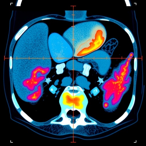
For decades, body fat has been reduced to a handful of numbers: body mass index, waist circumference and total fat percentage. These measurements are useful, but they can conceal a more complicated biological reality. Two people with similar body shapes may carry abdominal fat with very different internal structures and metabolic behaviors. A new study published in the International Journal of Obesity suggests that computed tomography, combined with advanced image analysis, can reveal those hidden differences in unprecedented detail, separating the characteristics of subcutaneous abdominal fat from those of visceral abdominal fat and exposing patterns linked to cardiovascular risk.
The research, led by Ana Rita Lopes, Inês Sousa, Fernando Nunes and colleagues, examines two major abdominal fat compartments. Subcutaneous abdominal fat, or SAF, lies directly beneath the skin, while visceral abdominal fat, or VAF, surrounds internal organs deep inside the abdomen. Although both are commonly grouped under the broad label of “body fat,” they are not biologically interchangeable. Visceral fat is more strongly associated with insulin resistance, chronic inflammation, abnormal blood lipids and atherosclerotic cardiovascular disease. Yet even within these compartments, fat tissue may differ in ways that conventional measurements cannot detect.
The investigators used computed tomography images to build radiomic profiles of SAF and VAF. Radiomics is a computational approach that converts medical images into large sets of quantitative features. Instead of measuring only the amount or thickness of fat, the method analyzes the internal appearance of tissue at the pixel or voxel level. These features can include intensity distribution, variation, spatial relationships, texture, uniformity and subtle patterns that are invisible to the human eye. In practical terms, radiomics treats a CT scan not simply as a picture, but as a source of biological data that may reflect tissue composition, cellular organization, fluid content, fibrosis and other disease-related processes.
This distinction is important because fat tissue is not a passive energy reservoir. Adipose tissue contains adipocytes, immune cells, blood vessels and connective-tissue structures, all of which can change under metabolic stress. Enlarged or dysfunctional fat cells may release inflammatory signaling molecules and alter the flow of fatty acids into the bloodstream. Visceral fat can also influence nearby organs through local inflammation and systemic endocrine effects. A radiomic signature may therefore act as an indirect fingerprint of tissue quality, capturing biological differences that are missed when researchers consider only how much fat is present.
The study identified clear differences between the radiomic profiles of subcutaneous and visceral abdominal fat, indicating that the two compartments possess distinct imaging phenotypes. It also found evidence of sexual dimorphism, meaning that the organization and characteristics of abdominal fat differed between women and men. This finding reinforces a growing body of research showing that adipose distribution is influenced by sex hormones, genetics, age, menopause, body composition and metabolic status. Women and men may accumulate fat in different locations and may experience different biological consequences from comparable quantities of adipose tissue. Treating body fat as a uniform tissue could therefore obscure clinically meaningful risk patterns.
Beyond sex-specific differences, the researchers identified radiomic signatures associated with a pro-atherogenic profile. Atherosclerosis develops through a complex interaction of lipid accumulation, vascular inflammation, immune responses and structural changes in the arterial wall. The term “pro-atherogenic” describes biological conditions that may favor this process, including patterns related to adverse lipid metabolism and inflammation. The study’s results suggest that the internal imaging characteristics of abdominal fat may contain information connected to cardiovascular vulnerability, even when standard body-size measurements provide a less concerning picture.
The potential clinical significance lies in the incremental value of these imaging features. Traditional cardiovascular risk assessment commonly combines age, blood pressure, cholesterol levels, smoking status, diabetes and other established variables. Measures such as waist circumference and body mass index may add information, but they do not directly describe the biological quality of adipose tissue. Radiomic analysis could eventually complement these tools by identifying patients whose abdominal fat appears metabolically or vascularly hazardous despite apparently ordinary anthropometric measurements. It might also help distinguish patients with similar visceral-fat volumes but different levels of tissue-related risk.
The work does not suggest that CT radiomics is ready to replace established clinical assessments, nor does it show that a particular radiomic pattern directly causes cardiovascular disease. Radiomic features can be influenced by scanner type, image acquisition settings, reconstruction algorithms, segmentation methods and the software used to extract measurements. Reliable clinical implementation would require standardized protocols, external validation in diverse populations and prospective studies showing that the signatures improve outcomes or change treatment decisions. Radiation exposure and the cost of CT also mean that the technique will need to be used selectively, potentially taking advantage of scans already obtained for other medical reasons.
Even with those limitations, the study points toward a broader transformation in how obesity and cardiovascular risk may be evaluated. Instead of asking only how much fat a person carries, clinicians and researchers may increasingly ask where that fat is located, how it is organized and what biological signals it appears to encode. The combination of anatomical imaging, radiomics and clinical data could make it possible to construct more individualized risk profiles, while sex-specific analysis may prevent important differences from being hidden inside population averages.
The findings place abdominal adipose tissue at the center of a new precision-medicine conversation. CT radiomics cannot yet provide a simple answer to who will develop a heart attack or stroke, but it may help reveal why conventional measures sometimes fail to distinguish low-risk from high-risk individuals. By mapping the hidden texture of subcutaneous and visceral fat, Lopes and colleagues offer evidence that the body’s fat stores are not merely quantities to be measured, but complex organs whose imaging signatures may carry clues about cardiovascular health. As larger studies test these signatures across populations and clinical settings, the technology could help turn routine medical images into more sensitive tools for detecting pro-atherogenic biology before overt disease appears.
Cite Scienmag News
Ophelia Keating. (August 20, 2026). Sex Differences and Atherosclerosis-Linked CT Patterns in Abdominal Fat. Scienmag. https://scienmag.com/sex-differences-and-atherosclerosis-linked-ct-patterns-in-abdominal-fat/
Ophelia Keating. "Sex Differences and Atherosclerosis-Linked CT Patterns in Abdominal Fat." Scienmag, 20 August 2026, https://scienmag.com/sex-differences-and-atherosclerosis-linked-ct-patterns-in-abdominal-fat/. Accessed 3 September 2026.
Ophelia Keating. "Sex Differences and Atherosclerosis-Linked CT Patterns in Abdominal Fat." Scienmag. August 20, 2026. https://scienmag.com/sex-differences-and-atherosclerosis-linked-ct-patterns-in-abdominal-fat/


