


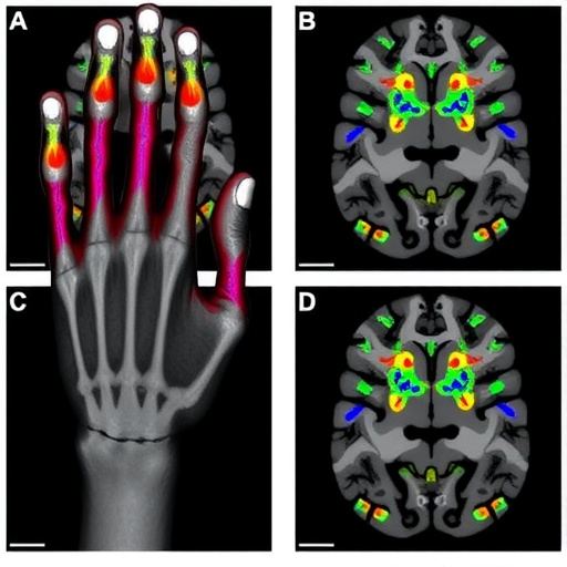
A nerve in the hand may be only a few millimeters wide, yet severing it can alter the brain’s detailed map of the fingers, according to a new study published in Nature Human Behaviour. The research by Michael Weber, Alexander Marshall, Raluca Timircan and colleagues provides fresh evidence that the primary somatosensory cortex is not organized as a permanently fixed anatomical chart. Instead, the cortical representation of the digits appears to depend on the sensory signals continually arriving from the body. When those signals are interrupted, the brain’s map can change in ways that challenge long-standing assumptions about how sharply the fingers are separated in the cortex.
The primary somatosensory cortex, or S1, lies along the postcentral gyrus of the brain and processes touch, pressure, vibration, temperature and information about the position of body parts. It is often described as a sensory homunculus: a distorted map in which neighboring areas of the body occupy neighboring regions of cortical tissue. The hand and fingers receive disproportionately large representation because they are essential for fine manipulation and contain dense populations of sensory receptors. Within the hand region of S1, individual digits have traditionally been thought to form orderly, partially overlapping bands. This arrangement has been used to explain the brain’s ability to distinguish one fingertip from another with extraordinary precision.
The new findings focus on what happens when one of the nerves carrying information from the hand is cut. A nerve injury of this kind does not simply remove sensation from the skin. It also deprives the brain of the patterned stream of electrical activity that normally identifies where a stimulus has occurred and which finger has been touched. Sensory neurons in the peripheral nervous system convert mechanical pressure or movement into impulses, which travel through the spinal cord and thalamus before reaching S1. When a nerve is interrupted, that communication pathway is disrupted. The study shows that the consequences extend beyond a silent patch of cortex: the structure of digit representations themselves can be reorganized.
This result is important because it bears on a decades-old debate in neuroscience. Some earlier experiments suggested that the brain contains stable, pre-existing cortical territories for each finger, with injury causing neighboring representations to expand into the deprived region. Other work has argued that apparent remapping may reflect changes in the way neurons respond, rather than a wholesale physical takeover of one cortical area by another. By examining digit maps after nerve injury, the researchers add evidence for a more nuanced model. The cortex remains anatomically continuous, but the functional boundaries between the fingers are flexible and can be reshaped when normal sensory input disappears.
The distinction between anatomical stability and functional reorganization is central to understanding the discovery. Neurons in S1 do not respond to only one perfectly defined point on the hand. Most have receptive fields, areas of the body from which stimulation can influence their activity. Receptive fields can overlap, and their size and sensitivity are affected by context, attention, learning and injury. A nerve lesion changes the statistical pattern of activity reaching the cortex, potentially altering the balance between signals from adjacent digits, the strength of connections within local cortical circuits and the influence of feedback from higher brain regions. Together, these changes can make the cortical map less like a set of rigid borders and more like a dynamic landscape.
The study also speaks to the sensory confusion many people experience after peripheral nerve damage. Patients may lose touch or feel numbness in specific parts of the hand, but they can also report imprecise localization, abnormal tingling or sensations that seem to spread across neighboring fingers. Such symptoms may arise because the brain is attempting to interpret incomplete or distorted input using an internal model built from previous experience. If signals that once arrived through one nerve are weakened or absent, activity from adjacent pathways may become relatively more influential. The resulting percepts do not necessarily correspond neatly to the original skin territory, even when the injury itself is clearly localized.
For researchers developing treatments, the findings may have practical implications. Nerve repair and rehabilitation are not only matters of reconnecting damaged fibers in the arm or hand. The central nervous system must also learn to interpret the signals that eventually return. After surgical repair, regenerating axons may reconnect imperfectly, and their signals may arrive with altered timing or at unusual cortical locations. Targeted sensory training, tactile discrimination exercises, virtual-reality interfaces and brain-computer technologies could help refine these representations. A clearer understanding of how digit maps change may allow clinicians to design rehabilitation programs that encourage useful reorganization rather than leaving the brain to adapt to noisy or ambiguous input.
The work also has significance beyond hand injuries. It contributes to a broader picture of the adult brain as plastic but constrained. Plasticity does not mean that any cortical area can instantly become any other function. The existing architecture, local connections, developmental history and ongoing sensory experience all impose limits. Yet the organization of the hand is evidently responsive to changes in peripheral input, demonstrating that even highly specialized sensory maps can be modified in adulthood. This flexibility may help the nervous system cope with injury, but it may also contribute to chronic pain, phantom sensations and difficulties with prosthetic devices when the new organization is maladaptive.
By showing that cutting a hand nerve can alter the organization of digit maps in S1, Weber and colleagues place peripheral nerve injury at the center of a question that reaches into the foundations of perception: does the brain represent the body as it is, or as it expects the body to be based on incoming signals? The evidence favors an interaction between both. The body provides the wiring and the sensory input, while the brain continually recalibrates its interpretation. A severed nerve therefore does more than interrupt communication between skin and cortex. It changes the information on which the brain’s map is built, revealing that the apparent boundaries between our fingers are not merely drawn into the cortex once and for all, but are maintained by experience.
Cite Scienmag News
Cassandra Pierce. (August 13, 2026). Severing a hand nerve reorganizes finger maps in primary somatosensory cortex. Scienmag. https://scienmag.com/severing-a-hand-nerve-reorganizes-finger-maps-in-primary-somatosensory-cortex/
Cassandra Pierce. "Severing a hand nerve reorganizes finger maps in primary somatosensory cortex." Scienmag, 13 August 2026, https://scienmag.com/severing-a-hand-nerve-reorganizes-finger-maps-in-primary-somatosensory-cortex/. Accessed 3 September 2026.
Cassandra Pierce. "Severing a hand nerve reorganizes finger maps in primary somatosensory cortex." Scienmag. August 13, 2026. https://scienmag.com/severing-a-hand-nerve-reorganizes-finger-maps-in-primary-somatosensory-cortex/


