


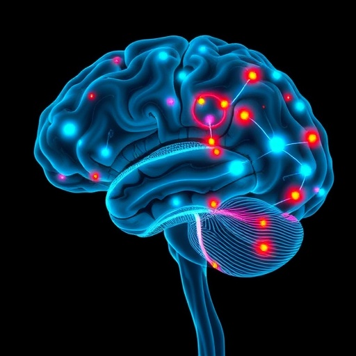
Adolescents with bipolar disorder and major depressive disorder show measurably different patterns of functional brain network organization, according to a new resting-state functional magnetic resonance imaging study published in BMC Psychiatry. The findings suggest that the topology of specific nodes within the default mode, visual, and prefrontal systems may hold diagnostic information capable of distinguishing the two conditions, which are frequently confused in clinical practice during adolescence.
The study, led by Yitong Liu, Yue Zhang, and Cai Li of the First Affiliated Hospital of Zhengzhou University together with colleagues at the Suzhou Mental Health Center, addressed a persistent problem in child and adolescent psychiatry: bipolar disorder in young people often presents initially with depressive symptoms, and the overlap in clinical features, combined with the absence of objective diagnostic markers, frequently results in bipolar disorder being misdiagnosed as major depressive disorder. Such misdiagnosis can carry significant consequences, since the two conditions may call for different treatment strategies. Direct comparisons of functional brain network topology between adolescents with the two disorders had remained limited, particularly with respect to how network alterations relate to specific clinical symptom dimensions.
To address this gap, the research team recruited a total of 134 participants: 55 adolescents with major depressive disorder, 35 with bipolar disorder, and 44 healthy controls. The researchers hypothesized that the two patient groups would exhibit distinct patterns of functional brain network organization, and that these patterns would be associated with specific clinical symptoms. All participants underwent resting-state functional MRI, a technique that measures spontaneous brain activity while participants lie awake but at rest, allowing researchers to map the functional connections that organize the brain into large-scale networks.
The analytical approach rested on graph theory, a mathematical framework in which brain regions are treated as nodes and functional connections between them as edges. From this network representation, the team computed several nodal metrics, including degree centrality, clustering coefficient, nodal efficiency, nodal local efficiency, and shortest path length. These measures capture different aspects of how well connected, how locally clustered, and how efficiently integrated each individual brain region is within the wider network. By comparing these metrics across the three groups, the investigators could identify which nodes showed disorder-specific alterations rather than changes shared across all mood disorders.
The comparisons yielded a differentiated picture. Relative to healthy controls, adolescents with major depressive disorder showed reduced nodal connectivity and efficiency in visual cortical regions, pointing to alterations in sensory-processing areas that have increasingly attracted attention in mood disorder research. More strikingly, when the two patient groups were compared directly, adolescents with bipolar disorder exhibited higher nodal metrics in specific nodes within the default mode network and prefrontal regions relative to those with major depressive disorder. The default mode network, which is most active during rest and self-referential thought, has been repeatedly implicated in affective disorders, while prefrontal regions are central to emotional regulation and cognitive control.
Beyond group differences, the team examined whether the altered nodal metrics were related to the clinical presentations of the patients. Correlation analyses revealed that clustering coefficients of the right dorsolateral superior frontal gyrus and the right orbital superior frontal gyrus were positively associated with performance on the attention/vigilance domain, suggesting that the local organization of these prefrontal nodes relates to a core cognitive function often impaired in affective illness. In a dissociable pattern, the clustering coefficient of the right cuneus, a region of the visual cortex, was associated with depressive and anxiety symptoms. These relationships were statistically significant at p < 0.05, and they link network topology not merely to diagnoses but to transdiagnostic symptom dimensions: cognition on one hand and mood and anxiety on the other.
The clinical relevance of these topological differences was tested directly using machine learning. The researchers constructed support vector machine (SVM) classifiers using the significantly altered nodal metrics as classification features, with the goal of distinguishing adolescents with bipolar disorder from those with major depressive disorder. Model performance was evaluated using a nested cross-validation framework, a rigorous design in which feature selection and hyperparameter tuning are performed within inner loops of the cross-validation to avoid optimistic bias in the performance estimates. A linear-kernel SVM achieved a mean classification accuracy of 78.5 percent, a balanced accuracy of 74.0 percent, and an area under the receiver operating characteristic curve (AUC) of 0.739. While such figures fall short of the levels needed for stand-alone clinical diagnosis, they indicate that nodal topological features carry genuine information about which disorder an adolescent is experiencing—information that is not currently available from any objective test.
The researchers also attended to the methodological details that can confound resting-state fMRI studies, particularly in adolescent populations where head motion is a common concern. The supplementary and analytic framework referenced standard quality-control measures, including framewise displacement as a motion metric and consideration of global signal regression, and group comparisons were carried out using analysis of covariance with appropriate post hoc testing. Clinical characterization drew on well-established instruments, including the 24-item Hamilton Depression Rating Scale, the Hamilton Anxiety Rating Scale, the Young Mania Rating Scale, the Pittsburgh Sleep Quality Index, and the MATRICS Consensus Cognitive Battery, ensuring that the clinical correlations were anchored in validated assessments of mood, anxiety, sleep, and cognition.
The study was approved by the Ethics Committee of the First Affiliated Hospital of Zhengzhou University, and written informed consent was obtained from all participants and their legal guardians, with procedures conducted in accordance with the Declaration of Helsinki. The work was supported by the National Natural Science Foundation of China and by several Henan provincial research programs, reflecting a broader investment in precision approaches to psychiatric diagnosis in China. The article was published open access, with a preprint-style early version shared to provide faster access to the peer-reviewed findings.
Like all studies of this kind, the work carries limitations that temper interpretation. The sample sizes, while respectable for clinical neuroimaging—particularly the 35 adolescents with bipolar disorder—moderate the statistical power available for detecting subtle network differences and for training robust classifiers. The cross-sectional design cannot determine whether the observed topological differences are stable traits, state-dependent features tied to current mood episode, or consequences of medication or illness course, none of which can be fully disentangled in a single scanning session. The reported classification performance, while promising, would need to be replicated in independent cohorts before any translation toward clinical decision support could be contemplated, and the modest AUC of 0.739 places the model in the range of a useful adjunct rather than a definitive test.
Nevertheless, the implications of the findings are substantial. First, they provide converging evidence that adolescent bipolar disorder and major depressive disorder, despite their symptomatic overlap in depressive phases, are distinguishable at the level of functional brain network architecture. The elevation of nodal metrics in default mode and prefrontal nodes in bipolar disorder, set against reductions in visual cortical connectivity and efficiency in depression, suggests partly distinct neural mechanisms rather than a single continuum of mood pathology. Second, the dissociation between prefrontal clustering coefficients linked to attention and vigilance and visual-node clustering linked to depressive and anxious symptoms supports a dimensional view in which specific network features map onto specific symptom domains—a framework consistent with contemporary efforts such as the Research Domain Criteria, which seek to anchor psychopathology in brain-based dimensions.
Third, and perhaps most practically, the successful use of nodal topological features to classify patients above chance demonstrates a feasible pipeline for developing imaging biomarkers. The features involved are computable from standard resting-state fMRI acquisition, which is non-invasive, widely available, and already used in research and some clinical settings. If future studies confirm and refine these classifiers, resting-state network metrics could eventually supplement clinical interview in the difficult early differentiation of bipolar disorder from unipolar depression in adolescents—a differentiation that currently relies entirely on clinical judgment, often delayed until a manic episode emerges.
The authors frame their conclusions cautiously, emphasizing that adolescents with the two disorders exhibited distinct patterns of nodal functional brain network organization particularly within the default mode, visual, and prefrontal systems, that altered topology was associated with cognitive and affective symptom dimensions, and that the SVM analyses suggest these features contain information relevant to differentiating the disorders. They position the work as providing further insight into the neural mechanisms underlying adolescent affective disorders rather than as an immediately deployable diagnostic tool.
For clinicians and researchers, the study adds a node-level perspective to a growing literature on large-scale network dysfunction in youth mood disorders. Previous work has often focused on whole-network summary measures or on seed-based connectivity between particular region pairs; by examining nodal metrics across the whole brain and linking them to fine-grained clinical measures, this study identifies specific anatomical loci—the right dorsolateral and orbital superior frontal gyri, the right cuneus, and default mode nodes—where topology tracks diagnosis and symptoms. Future longitudinal research, ideally following high-risk adolescents over time and incorporating treatment response, will be needed to determine whether these topological signatures precede illness onset, predict conversion from depressive to bipolar presentations, or change with effective intervention. In the interim, the study stands as a methodologically careful demonstration that the architecture of the resting brain differs in measurable and clinically meaningful ways between adolescents with bipolar disorder and those with major depressive disorder, bringing the field a step closer to objective, biology-informed diagnosis of the most diagnostically challenging period in mood disorder medicine.
Cite Scienmag News
Glenn Wilkins. (August 31, 2026). Differential nodal topology in resting-state networks as a potential imaging marker for adolescent bipolar and depressive disorders. Scienmag. https://scienmag.com/differential-nodal-topology-in-resting-state-networks-as-a-potential-imaging-marker-for-adolescent-bipolar-and-depressive-disorders/
Glenn Wilkins. "Differential nodal topology in resting-state networks as a potential imaging marker for adolescent bipolar and depressive disorders." Scienmag, 31 August 2026, https://scienmag.com/differential-nodal-topology-in-resting-state-networks-as-a-potential-imaging-marker-for-adolescent-bipolar-and-depressive-disorders/. Accessed 3 September 2026.
Glenn Wilkins. "Differential nodal topology in resting-state networks as a potential imaging marker for adolescent bipolar and depressive disorders." Scienmag. August 31, 2026. https://scienmag.com/differential-nodal-topology-in-resting-state-networks-as-a-potential-imaging-marker-for-adolescent-bipolar-and-depressive-disorders/


