


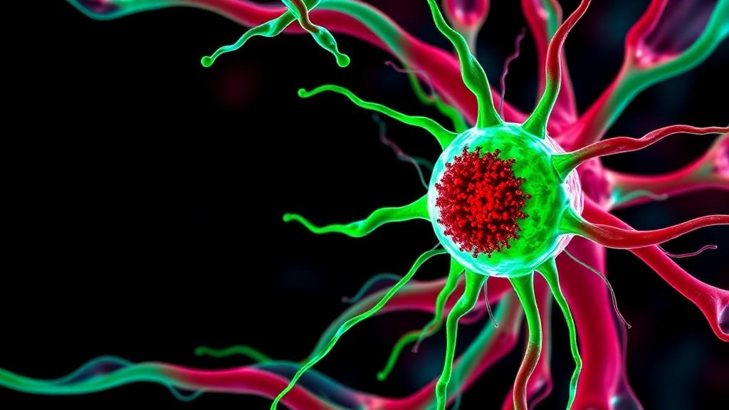
Scientists at Weill Cornell Medicine have identified a previously unknown cell type in the zebrafish heart that appears to act as a built-in brake on blood vessel growth, a finding that could reshape how researchers think about vascular development and, ultimately, the regeneration of damaged human heart tissue. The discovery, published on August 20 in Nature Communications, centers on a population of perivascular cells that wrap around the coronary vessels supplying oxygen to the cardiac muscle. Rather than simply supporting the growing vasculature, these cells appear to actively regulate it, producing molecular signals that rein in vessel expansion once the network has reached the appropriate scale. The work emerges from a laboratory whose long-term ambition is one of the most sought-after goals in cardiovascular biology: rebuilding a heart that has been damaged by disease or injury.
The heart is an organ with an unforgiving relationship to its blood supply. Cardiac muscle works continuously and demands a constant delivery of oxygen, which means the coronary vasculature must grow in careful proportion to the muscle it serves. Too little vessel growth starves the tissue; too much creates a disorganized, inefficient network. During development, and again after injury, the heart must therefore solve a problem that engineers would recognize immediately: building the right amount of blood vessel, and knowing precisely when to stop. The new study suggests that the zebrafish heart solves this problem with a dedicated cellular control system, one that had gone unnoticed until now because it only becomes prominent under specific conditions of growth and repair.
The research is rooted in a question that has animated the field for years. Zebrafish possess a remarkable ability that mammals largely lack: after significant injury to the heart, they can regenerate the damaged muscle almost completely, restoring function rather than forming a permanent scar. Understanding how they accomplish this has become a major strategy for identifying the regenerative programs that might, in principle, be reawakened in human patients. Dr. Jingli Cao, associate professor of cell and developmental biology at Weill Cornell Medicine and a member of its Cardiovascular Research Institute, has spent much of his career tracing this process. As a postdoctoral fellow at Duke University, he determined that the epicardium, the thin layer of cells that envelops the heart, provides both the signals and the cells needed to regenerate heart tissue in zebrafish.
The epicardium has long been viewed as more than passive packaging. In response to injury, it activates a specific population of progenitor cells that give rise to the various cell types involved in regenerating heart muscle and coronary blood vessels. But a deeper puzzle remained: what directs these precursor cells to specialize into one fate or another? To answer it, Dr. Cao’s team, including graduate student Björn Perder and postdoctoral fellow Dr. Yu Xia, catalogued the genes that switch on in these cells when the heart is injured. Among the activated genes they identified a master regulator called scxa, a transcription factor that steers a subset of epicardial progenitor cells toward a fate that had never been characterized before.
That fate turned out to be a novel perivascular cell type. Once specified by scxa, these cells migrate to the coronary vessels and surround them, taking up a position that immediately suggested a regulatory role. Perivascular cells in general are known to modulate vessel stability, maturation, and remodeling, so the location of the new cell type was a strong clue. The team found that these cells express a particular type of collagen, and that this collagen does more than provide structural support. The cells process the collagen molecule to release a small protein fragment, a derived signal that ultimately curbs further vessel expansion. In other words, the very cells that accompany the growing vasculature carry within them the molecular message that tells the network when enough is enough.
Dr. Cao framed the significance of this arrangement in practical terms. During development, or when the heart is damaged, the organism needs to build the right amount of blood vessel, and it needs to know when to stop, he noted. The newly discovered system provides that layer of control. This kind of negative feedback is a recurring theme in biology, but identifying the specific cell type and the specific signaling molecule responsible in the heart gives researchers a concrete handle on a process that has been difficult to study. It also suggests that disorders of vascular growth in the heart, whether insufficient vessel formation after injury or excessive, disorganized growth, might be traceable to failures of this regulatory circuit.
Perhaps the most intriguing aspect of the study is what it reveals about timing. The researchers had previously shown that when regions of the heart experience low oxygen levels, a condition known as hypoxia, signals from the epicardium coordinate the growth of heart muscle and coronary vessels so that the two develop in step with each other. The new findings illuminate a plausible mechanism for how this coordination happens. Low oxygen appears to act as an environmental cue that temporarily activates scxa, which in turn directs a subset of epicardial progenitors toward the perivascular fate. These cells then surround the coronary vessels and may help regulate their development and remodeling. Hypoxia, in this model, is not merely a stress signal but an instruction: it tells the epicardium that the tissue is outpacing its blood supply and that vascular support cells are needed now.
This oxygen-sensing logic has an elegant developmental rationale. As heart muscle proliferates, the growing tissue inevitably pushes local oxygen levels down, because diffusion cannot keep pace with expansion. By using hypoxia as the trigger for scxa activation, the heart ensures that vessel-supporting cells are generated exactly where and when the muscle is growing fastest. The same mechanism likely operates after injury, when the damaged tissue becomes hypoxic and the epicardium mounts its regenerative response. If the system works as the study suggests, the perivascular cells and their collagen-derived braking signal would form a self-correcting loop: hypoxia recruits the cells, the cells support vessel growth, and the vessels restore oxygen delivery, at which point the signal to expand fades.
The translational implications are considerable, though the researchers are careful about the distance between zebrafish and humans. Humans do carry a related gene, SCX, but no one yet knows whether human epicardial cells use it in the same way as their zebrafish counterparts. There is a cautionary wrinkle: after an injury such as a heart attack in mammals, SCX is switched on in cardiac fibroblasts, the cells that promote scar tissue formation. In mammals, then, the same regulatory gene appears to be associated with fibrosis rather than regeneration. Dr. Cao suggests that in the future it may be possible to use SCX to reactivate human epicardial cells and steer them toward generating the cells and signals that promote heart regeneration, redirecting the gene’s activity away from scarring and toward repair.
Toward that end, Dr. Cao’s laboratory is working with cultured human epicardial cells and cardiac organoids, laboratory-grown miniature tissue models, to develop ways of using activated cells to repair damaged regions of the heart. The vision is a kind of biological bandage: a living material that could be applied to an injured site and induce controlled growth of both heart muscle and vessels, rebuilding the tissue in a coordinated fashion rather than leaving a non-contractile scar. Dr. Cao is quick to temper expectations, noting that he does not believe a single magic factor controls regeneration. But, as he put it, the team has discovered one more factor, one more mechanism that could eventually contribute to turning on epicardial cells and repairing human hearts. The work was supported by the New York State Stem Cell Science program, the Louis and Rachel Rudin Foundation, the Belgian American Educational Foundation, the American Heart Association, and the National Institutes of Health, reflecting the broad institutional bet that the zebrafish heart still has many secrets worth stealing.
Subject of Research: Discovery of a novel scxa-specified perivascular cell type that regulates coronary vascular development in the regenerating zebrafish heart
Article Title: Newly discovered cell helps shape blood vessel growth in the heart
Article References: Newly discovered cell helps shape blood vessel growth in the heart. (n.d.). Original publication
Image Credits: AI Generated
DOI: Not provided
Keywords: zebrafish, heart regeneration, epicardium, perivascular cells, coronary vessels, scxa, hypoxia, collagen, Weill Cornell Medicine, Nature Communications, cardiac progenitor cells, vascular development
Cite Scienmag News
Drew Townsend. (October 3, 2026). Scientists discover a novel heart cell that tells blood vessels when to stop growing. Scienmag. https://scienmag.com/scientists-discover-a-novel-heart-cell-that-tells-blood-vessels-when-to-stop-growing/
Drew Townsend. "Scientists discover a novel heart cell that tells blood vessels when to stop growing." Scienmag, 3 October 2026, https://scienmag.com/scientists-discover-a-novel-heart-cell-that-tells-blood-vessels-when-to-stop-growing/. Accessed 4 October 2026.
Drew Townsend. "Scientists discover a novel heart cell that tells blood vessels when to stop growing." Scienmag. October 3, 2026. https://scienmag.com/scientists-discover-a-novel-heart-cell-that-tells-blood-vessels-when-to-stop-growing/


