


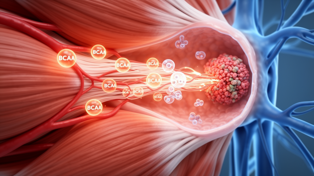
Cancer can reshape how skeletal and cardiac muscles handle branched-chain amino acids (BCAAs), the essential nutrients leucine, isoleucine and valine, according to a review comparing normal metabolism with changes seen in tumor-bearing models and patients. The synthesis finds that these amino acids have two major roles: they supply building blocks for proteins and signal through nutrient-sensing pathways, while their breakdown provides carbon compounds that enter mitochondrial energy production. Their fate is not uniform, however. Skeletal muscle is a major site of BCAA transamination and oxidation across the body, whereas healthy cardiac muscle obtains less than 2% of its ATP from BCAA and depends mainly on glucose and fatty acids. Cancer adds another layer because different malignancies either increase or suppress amino-acid catabolism in ways that may support tumor growth while depriving or stressing host muscle. The review also separates direct observations in cancer-cachexia models from cardiac mechanisms extrapolated from heart-failure research.
The biochemical starting point is the enzyme branched-chain aminotransferase, which transfers the amino group from each BCAA to alpha-ketoglutarate, producing glutamate and the corresponding branched-chain alpha-keto acid. This reversible reaction operates close to equilibrium, so its net direction depends on what happens to the keto acids afterward. Mitochondrial BCAT2 is expressed in skeletal muscle and other tissues, whereas cytosolic BCAT1 is concentrated mainly in the central nervous system but appears in some cancers. A second complex, the branched-chain alpha-keto acid dehydrogenase complex, or BCKDH, then oxidatively decarboxylates those keto acids. Its activity is controlled by a balance between BCKDH kinase, which phosphorylates and inhibits the complex, and protein phosphatase 2C, encoded by PPM1K, which dephosphorylates and reactivates it. The products differ according to the original amino acid: leucine yields acetyl-CoA and acetoacetate, isoleucine yields acetyl-CoA and succinyl-CoA, and valine yields succinyl-CoA. These products can replenish the tricarboxylic acid cycle and support mitochondrial energy production.
In skeletal muscle, BCAA metabolism is best understood as a flexible partition between protein turnover and oxidative disposal. After feeding, many dietary BCAAs are retained for synthesis and replacement of contractile proteins. During fasting, prolonged exercise or systemic stress, oxidative flux rises, shifting amino acids away from protein incorporation and toward energy production and nitrogen turnover. BCAT2 and BCKDH allow this adjustment while limiting the buildup of potentially harmful keto-acid intermediates. Leucine also has a signaling role: when total essential-amino-acid availability and the hormonal setting are permissive, it can activate mTORC1-dependent translation. This combination of substrate delivery and nutrient signaling explains why BCAA abundance alone does not fully determine whether muscle protein synthesis rises.
Cardiac muscle shares the same catalytic machinery but applies it under different constraints. Because the heart contracts continuously, it normally relies overwhelmingly on fatty acids and glucose, and its total contribution to whole-body BCAA oxidation is small relative to that of skeletal muscle and adipose tissue. Tightly controlled BCAA flux supports renewal of contractile and mitochondrial proteins, but it also prevents BCAA-derived keto acids from accumulating and disturbing mitochondrial respiration and redox balance. In pressure-overloaded mouse hearts and human dilated cardiomyopathy, genes encoding BCAT2, BCKDH subunits and PPM1K can be downregulated, leading to inhibitory phosphorylation of BCKDH and accumulation of keto acids. Those intermediates inhibited complex I-dependent respiration and increased superoxide production in isolated mitochondria. This mechanism is biologically plausible for cancer-related cardiac dysfunction, but it has not yet been established directly in a tumor-bearing host.
Tumors themselves exploit BCAA metabolism in several different ways. Many cancers reduce the breakdown of BCAAs, allowing intracellular pools to remain available for protein synthesis and sustained mTORC1 signaling, particularly when mitogenic signals and nutrients are both present. A pan-cancer analysis cited by the review estimated that about 70% of malignancies suppress BCAA metabolism, although this pattern is not universal. In head and neck squamous cell carcinoma, melanoma, breast cancer and some gliomas, BCAT1 is instead elevated and associated with proliferation, migration or other tumor-promoting activity. The review therefore cautions against treating BCAA metabolism as a single cancer hallmark with one direction. Tumor origin, molecular background and stage all matter, and findings from one malignancy should not automatically be generalized to another.
Cancer cachexia connects tumor metabolic reprogramming to a multi-organ syndrome in which inflammation, endocrine disturbance, mitochondrial dysfunction and impaired anabolic signaling drive wasting of muscle and other tissues. In skeletal muscle, cytokines including interleukin-6, tumor necrosis factor-alpha and interferon-gamma activate pathways such as NF-kappaB and STAT3, increasing the muscle-specific ubiquitin ligases atrogin-1 and MuRF1. Autophagy-lysosomal pathways may also become more active. At the same time, the PI3K-Akt-mTOR network becomes less responsive to nutrients and growth signals, so protein degradation is not balanced by adequate replacement. Mitochondrial respiration weakens, ATP production falls and reactive oxygen species increase, while mitophagy rises alongside reduced mitochondrial biogenesis. BCAA handling sits within this network rather than operating independently: supplying more amino acids cannot by itself correct impaired transport, inflammatory signaling or mitochondrial quality control.
Experimental mouse models show that this relationship can be causal in at least some contexts. In mice bearing Lewis lung carcinoma, muscle-specific deletion of Bckdk, which relieves inhibition of BCKDH, lowered BCAA concentrations in muscle and serum, reduced mTOR signaling, accelerated cachexia and shortened survival. A BCAA-enriched diet extended survival in those knockout animals but did not do so in tumor-bearing wild-type mice. In the C26 colon carcinoma model, concentrations of all three BCAAs fell in skeletal muscle, while tumor BCAA content and BCKDH activity increased later in disease progression. These opposite movements illustrate why plasma BCAA concentrations are a weak proxy for any single tissue: circulating values represent the net result of opposing changes among tumor, muscle, kidney and other organs. Transporters also alter intracellular availability independently of the bloodstream, with reduced LAT1 expression observed in peripheral tissues of C26-bearing mice.
The principal cancer examples reveal sharply different tumor strategies. Non-small cell lung cancer avidly takes up circulating BCAAs and uses BCAA-derived nitrogen for biosynthesis, including nucleotide production. Pancreatic ductal adenocarcinoma, by contrast, takes up less BCAA than non-small cell lung cancer in autochthonous mouse models and relies more on alternative nitrogen sources, although some experimental PDAC systems show increased BCAA use. Patients with pancreatic cancer can have elevated circulating BCAAs two to five years before diagnosis, but this temporal association reflects altered whole-body metabolism rather than proving a particular tumor or muscle mechanism. Lung cachexia also adds respiratory compromise and disuse to systemic inflammation, creating a cycle in which reduced activity lowers muscle loading while catabolic signaling rises. For PDAC, experimental evidence highlights a tumor-derived interleukin-6 trans-signaling loop involving adipose tissue and skeletal muscle, with disruption of tumor interleukin-6 reducing lipid accumulation and atrophy in orthotopic and patient-derived mouse models.
Cardiac involvement has been documented more clearly than the proposed BCAA-specific mechanism behind it. C26 tumor-bearing mice showed increased cardiac MuRF1 and atrogin-1 expression, greater protein ubiquitination, and reductions of 43% in myosin heavy chain and 58% in troponin I. Pancreatic and ovarian cancer models have shown cardiac atrophy, contractile dysfunction and disturbed mitochondrial energetics, while patients with advanced cancer can display reduced left ventricular mass, chamber volumes and stroke volume despite preserved ejection fraction. In pancreatic cancer models, myocardial atrophy has also been associated with increased MAO-A, atrogin-1 and interleukin-1-beta, although inhibition of MAO-A with harmine did not assess cardiac function in that study. Notably, measurements of myocardial BCKDH activity, PPM1K expression and intramyocardial keto-acid content have not been reported in cancer-cachexia models. The strongest mechanistic claims connecting impaired cardiac BCAA catabolism to cancer therefore remain extrapolations from heart failure.
The review identifies several priorities before BCAA-directed strategies can be translated. Tumor heterogeneity means interventions will not have uniform effects: inhibiting BCKDK may restrain a tumor that depends on suppressed BCAA catabolism, yet activating the same pathway in host muscle can reduce mTOR signaling and worsen wasting. The timing of treatment is equally important. Cachexia progresses from precachexia to cachexia and refractory stages, with anabolic responsiveness declining as irreversible wasting develops, making early intervention more plausible than late supplementation. Evidence is also constrained by treatment effects: FOLFOX and FOLFIRI can independently cause muscle loss, reduce mitochondrial content and alter LAT1, BCKDH activity and intracellular BCAAs in experimental systems, while anthracyclines can reduce cardiac mass. Future work should therefore measure tissue-specific BCAA flux, distinguish tumor actions from host responses, and test combinations that suppress inflammation and endoplasmic-reticulum stress while restoring mitochondrial quality control rather than treating amino-acid availability as a stand-alone solution.
Subject of Research: Cancer-driven changes in branched-chain amino acid metabolism in skeletal and cardiac muscle during cachexia
Article Title: Branched-chain amino acids in skeletal and cardiac muscles: a review of metabolic variations and cancer’s impact
Article References: Chrzanowski, J., Graboń, W., Buchajska, K., & Cudnoch-Jędrzejewska, A. (2026). Branched-chain amino acids in skeletal and cardiac muscles: a review of metabolic variations and cancer’s impact. Frontiers in Physiology, 17, 1943433. https://doi.org/10.3389/fphys.2026.1943433
Image Credits: AI Generated
DOI: 10.3389/fphys.2026.1943433
Keywords: branched-chain amino acids, cancer cachexia, skeletal muscle, cardiac muscle, tumor metabolism, BCAA catabolism, mTORC1 signaling, mitochondrial dysfunction, muscle wasting, BCKDH, Branched-chain, amino
Cite Scienmag News
Nathaniel Bowman. (October 22, 2026). Review maps cancer-linked BCAA rewiring in muscle. Scienmag. https://scienmag.com/review-maps-cancer-linked-bcaa-rewiring-in-muscle/
Nathaniel Bowman. "Review maps cancer-linked BCAA rewiring in muscle." Scienmag, 22 October 2026, https://scienmag.com/review-maps-cancer-linked-bcaa-rewiring-in-muscle/. Accessed 22 October 2026.
Nathaniel Bowman. "Review maps cancer-linked BCAA rewiring in muscle." Scienmag. October 22, 2026. https://scienmag.com/review-maps-cancer-linked-bcaa-rewiring-in-muscle/


