Often hailed as “the window to the soul,” the human eye has long fascinated scientists and philosophers alike. However, beyond its poetic allure, emerging research suggests that the eyes provide a vital window into the health of the brain. A groundbreaking new study spearheaded by the University of Florida’s biomedical engineering department has unveiled a transformative approach to predicting Alzheimer’s disease risk factors by analyzing retinal photographs with cutting-edge deep learning algorithms. This development promises to revolutionize early detection and intervention for one of the most devastating neurodegenerative diseases.
Traditionally, diagnosing Alzheimer’s disease has been a challenge due to its insidious onset and prolonged development over decades. Most diagnostic protocols focus on detecting the disease in its later stages, by which time significant, irreversible brain damage has already occurred. Recognizing this limitation, Dr. Ruogu Fang and her interdisciplinary team leveraged advances in artificial intelligence and biomedicine to explore the retina—a part of the central nervous system accessible via non-invasive imaging—as a potential biosensor for Alzheimer’s risk.
The study capitalized on a rich database of over 40,000 retinal images collected from the UK Biobank, a comprehensive health resource with longitudinal patient data. Retinal photography is commonly conducted during routine eye exams, diabetes management, glaucoma monitoring, and cataract assessments. By harnessing these widely available, inexpensive images, the research team circumvented the need for costly, invasive, or logistically complex diagnostic tools such as MRI or PET scans, thus holding promise for scalable, population-wide screening.
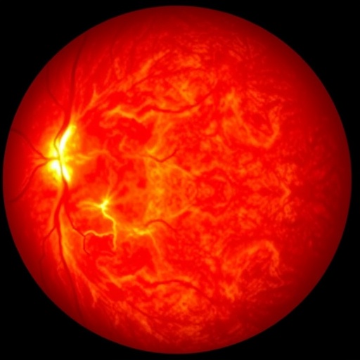
Employing deep learning, a subset of machine learning characterized by neural networks modeled after the human brain, the researchers developed an AI framework that scrutinized retinal morphology. This model was able to discern subtle variations in retinal arteries, veins, and the optic nerve head, features imperceptible to the human eye but correlated strongly with known risk factors for Alzheimer’s disease. Such morphological markers serve as proxies for neurovascular health, which is crucial given Alzheimer’s recognized association with vascular impairment.
One of the most striking revelations of this research was the model’s capacity not only to predict intrinsic biological traits such as sex and blood pressure but also modifiable lifestyle factors that influence Alzheimer’s risk. The AI effectively inferred behaviors like smoking, alcohol consumption, and even insomnia from retinal images alone. This objective measurement circumvents the notorious unreliability of self-reported lifestyle data, often plagued by bias and inaccuracies, thereby enriching risk assessment precision.
As Dr. Fang points out, retinal morphology functions less like a mere clinical questionnaire and more like an integrative biological sensor reflecting a cumulative lifetime burden of neurovascular and lifestyle insults. This conceptual shift underscores the retina’s role in encapsulating decades of physiological data, transforming retinal imaging into a powerful tool for capturing both present health and future vulnerability.
Furthermore, the implications extend beyond risk prediction. Identifying individuals with early signs of retinal changes could trigger timely clinical interventions long before cognitive symptoms manifest. Such preclinical detection enables the implementation of protective lifestyle adjustments, pharmacological therapies, or cognitive training programs designed to delay or mitigate Alzheimer’s progression.
The pioneering work built on previous findings from Fang’s group that demonstrated retinal images’ utility in detecting active Alzheimer’s disease cases. However, this new model pioneers the field by focusing on prodromal markers and risk factors instead of established dementia, bridging a critical gap in neurodegenerative diagnostics.
Importantly, the use of retinal photographs democratizes neurovascular health monitoring. Since retinal imaging is non-invasive, accessible, and affordable, it offers the prospect of integrating brain health assessment into routine eye care, thus expanding preventive neurology beyond specialized clinical settings.
This research also exemplifies the synergy between computational science and biomedical engineering. By combining sophisticated image analysis algorithms with large-scale patient data, it transcends traditional diagnostic constraints, marrying technology with medicine in a profoundly impactful manner.
While challenges remain, including the need for further validation across diverse populations and integration with other biomarkers, this study sets a new paradigm for early Alzheimer’s disease risk stratification. The possibility of preemptive interventions guided by retinal AI analysis heralds a future where the devastating trajectory of Alzheimer’s may be altered through timely and personalized healthcare.
In summary, the novel application of deep learning to retinal photographs opens an unprecedented window into brain health, heralding a new era of precision neurodiagnostics. It moves us closer to the long-sought goal of identifying Alzheimer’s risk decades in advance, fostering opportunities for early intervention and ultimately, better patient outcomes.
Subject of Research: People
Article Title: Prediction of Alzheimer’s disease risk factors from retinal images via deep learning: Development and validation of biologically relevant morphological associations in the UK Biobank
News Publication Date: 16-Jun-2026
Web References:
DOI: 10.1177/13872877261457650
Keywords
Alzheimer’s disease, Neurodegenerative diseases, Neurological disorders, Eye, Retina, Artificial intelligence, Image analysis, Deep learning, Biomedical engineering, Neurovascular integrity, Retinal biomarkers, Predictive modeling
