


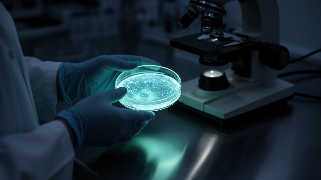
Bioluminescence is light produced by living organisms through chemical reactions, a phenomenon fundamentally different from incandescence, which arises from heat. A new review surveys this natural light from its chemical foundations to its ecological significance and its growing list of technological applications, noting that the capacity to emit light has evolved independently in many lineages, from deep-sea fish and dinoflagellates to fireflies, fungi, and bacteria. The core reaction involves a substrate called luciferin, which is oxidized by an enzyme called luciferase in the presence of oxygen.
The reaction produces an excited-state product, oxyluciferin, which releases a photon as it returns to the ground state. Ecologically, bioluminescence serves diverse roles. Fireflies use light patterns for communication and mating, with each species producing distinctive flash signatures.
Fungal bioluminescence produces a continuous green glow. Bacterial bioluminescence is regulated by quorum sensing, a mechanism in which bacteria produce and detect signaling molecules to coordinate gene expression according to population density. The lux operon governs the light-producing machinery.
Applications in basic research are extensive. In BRET, a bioluminescent donor transfers energy to a fluorescent acceptor, with the transfer efficiency indicating the distance between the two molecules. Quantum dots have been employed as acceptors in BRET configurations, taking advantage of their tunable optical properties.
Applied technology also draws on bioluminescence. ATP-based luminometers are widely used in the food industry for hygiene monitoring, providing rapid detection of residual organic contamination on surfaces. The review concludes that bioluminescence remains a vibrant field spanning basic research and applied technology.
The human fascination with living light is ancient. Descriptions of glowing organisms appear in writings attributed to Aristotle in the fourth century before the common era and to Pliny the Elder in the first century after it, long before any biochemical explanation existed. Modern surveys show that the trait appears in roughly 700 genera distributed across 16 major phyla, overwhelmingly in marine environments.
Notably, this broad phylogenetic spread does not follow a clear evolutionary pattern, suggesting that light production has arisen repeatedly and independently rather than from a single origin. In many animals, light is produced in specialized organs called photophores, whose activity can be governed by the nervous system. This neural control allows fish and squid to deploy their emissions contextually, for example to lure prey, deter predators, or signal potential mates.
In bacteria and fungi, by contrast, emission tends to be continuous because the cells constitutively contain all the molecular components needed for the reaction, and no regulatory trigger is required. Luminous fungi deserve particular attention. As of 2013, only 75 terrestrial fungal species were known to be bioluminescent, all within the order Agaricales and distributed across four evolutionary lineages: 52 species in the Mycenoidea group, five in Armillaria, twelve in Omphalotus, and two species originally described from Brazil, Gerronema viridilucens and Mycena lucentipes.
With the exception of some phytopathogens such as Armillaria mellea and Mycena citricolor, all known luminous fungi are saprotrophs, meaning they feed by absorbing organic substances from decaying matter. They are white-rot basidiomycetes whose mycelium secretes extracellular enzymes, including laccases and lignin and manganese peroxidases, capable of degrading lignin. The distribution of glowing tissue within these fungi is not uniform.
In some species both the mycelium and the entire fruiting body emit light, while in others the glow is restricted to the stipe, the pileus, or the lamellae. Regardless of location, all described species emit continuously rather than in flashes. The best-documented early example is Panellus stipticus, the bitter oyster, first described by the French mycologist Jean Bulliard in 1783 and given its current name in 1879.
Molecular phylogenetics places it close to the genus Mycena. Its greenish glow, strongest in damp, low-light habitats, is considered an evolutionary trait that may attract insects and help deter predators, contributing to survival. Another striking example is Neonothopanus gardneri, known in Brazil as the coconut flower, recorded in the Mata dos Cocais region of Maranhão and in Tocantins, Goiás, and Piauí.
Its fruiting bodies grow at the base of palms such as Attalea humilis, Attalea funifera, and Orbignya phalerata. Biochemical work published in 2017 confirmed that fungal light emission depends on four components: the substrate luciferin, a soluble NADPH- or NADH-dependent reductase, a membrane-bound oxygenase acting as luciferase, and molecular oxygen, consistent with a proposal made in 1960 by Airth and Foerster. The fungal chemistry was clarified in 2015, when the styrylpyrone 3-hydroxyhispidin was identified as the light-emitting substrate, derived from the precursor histidine through oxygenation catalyzed by a soluble NADPH-dependent hydroxylase.
The luciferin is then oxidized by an insoluble luciferase without requiring additional cofactors, producing green light at approximately 520 nanometers. Oxidation generates a high-energy endoperoxide intermediate that decarboxylates into fungal oxyluciferin and caffeylpyruvate; enzymatic hydrolysis of the latter yields caffeic acid, which is recycled through the histidine biosynthesis pathway via the styrylpyrone route. Because caffeic acid production ties into the Krebs cycle and the shikimic acid pathway, both shared by fungi and plants, this is the only eukaryotic bioluminescence system known to be genetically encodable, now documented in 125 species of luminous fungi.
Luciferin chemistry varies widely across the tree of life. Coelenterazine, found in jellyfish and corals, requires calcium ions and oxygen for its oxidation, and the color and intensity of the resulting emission depend on the molecular composition of the substrate and on environmental conditions. Vargulin operates in the ostracod crustacean Vargula hilgendorfii, where flavin adenine dinucleotide reductase and flavin mononucleotide reductase participate in the light-generating reduction reaction.
Biliverdin-based systems add further diversity. In most known systems, light arises from cleavage of a four-membered dioxanone ring, forming acyclic peroxide intermediates whose breakdown yields electronically excited products. The process is remarkably efficient, releasing most of its energy as light with little dissipation as heat.
Cofactors fine-tune these reactions. Calcium ions can activate luciferase in certain organisms by inducing a conformational change, and low calcium availability compromises light output. Magnesium ions similarly influence luciferase stability and activity, affecting oxidation efficiency.
Oxygen acts as the electron acceptor that forms the excited state from which a photon is emitted. Physics provides a quantitative framework for these processes. In Förster resonance energy transfer, the excited donor molecule passes its excess energy to an acceptor without emitting a photon.
Theodor Förster showed in 1948, using a fully quantum-mechanical treatment, that dipole-coupled transfer depends primarily on spectral overlap between donor emission and acceptor absorption and on intermolecular distance, with efficiency falling off as the inverse sixth power of the donor-acceptor separation. The Förster distance is defined as the separation at which transfer efficiency reaches fifty percent. Quantum yield, the ratio of photons emitted to photons absorbed, quantifies how completely absorbed energy is re-released as light.
In bioluminescence resonance energy transfer, the excited donor is generated biochemically rather than by external illumination, most commonly using Renilla luciferase oxidizing coelenterazine. Coupling this donor to semiconductor quantum dots, nanometer-scale crystals sometimes called artificial atoms, yields self-illuminating conjugates whose band gap, and therefore emission color, is inversely related to particle size. These conjugates combine adjustable emission, high brightness, and strong photostability, and have been used in nucleic acid hybridization assays, enzymatic activity detection, in vivo imaging, and biosensing.
Ecologically, predatory bioluminescence proceeds in three steps: light production, rapid emission, and prey capture, with flash frequency and intensity regulated biochemically to lure planktonic organisms. Some species also emit light under low nutrient availability in a process described as light respiration, in which energy released through luciferin oxidation may aid survival during food scarcity. Medical applications are advancing rapidly.
In rat models of gliosarcoma, cells transfected with luciferase-expressing plasmids allowed researchers to monitor photodynamic therapy in real time, showing increased cancer cell death when bioluminescence induction accompanied treatment. Bioluminescent quantification of HIV type 1 proved more sensitive than conventional phenotyping methods in settings lacking standard antibiograms. Firefly luciferase, favored for its higher quantum yield, has been used in D-luciferin oxygenation models to predict tumor development rates in vivo.
Combining bioluminescent proteins with positron emission tomography enables visualization of immune responses and drug distribution, although tissue imaging depth remains limited to roughly one to two centimeters and substrate administration is required before detection. In cosmetics, fluorescent makeup and skincare formulations inspired by luminous organisms are being explored, with some studies suggesting the underlying reaction may stimulate collagen production, potentially reducing wrinkles and age spots. Bioluminescent proteins also inspire novel pigments for experimental paints, plastics, and fabrics, though these lack the durability of traditional dyes.
In the food sector, ATP-bioluminescence testing validated organic contamination on restaurant workers' hands in Ouro Preto, Brazil, revealing loads above safe limits; after hygiene training, a fifty percent reduction was measured. Engineered biosensors emitting light in the presence of pathogens such as Salmonella and Escherichia coli enable rapid contamination screening, while modified lactic acid bacteria report pH changes and spoilage in fermented foods, and bioluminescent markers support ingredient traceability along supply chains.
Subject of Research: Bioluminescence mechanisms, ecological roles, and applications
Article Title: Unveiling bioluminescence: understanding mechanisms and exploring wide-ranging applications
Article References: Oliveira, M. D. D. A., Feitosa, C. M., Alves, P. E. S., Júnior, J. S. C., Lima, N. M., & Andrade, T. D. J. A. S. (2026). Unveiling bioluminescence: understanding mechanisms and exploring wide-ranging applications. Results in Chemistry, 31, Article 103946. https://doi.org/10.1016/j.rechem.2026.103946
Image Credits: AI Generated
DOI: 10.1016/j.rechem.2026.103946
Keywords: bioluminescence, luciferase, luciferin, BRET, biosensors, quorum sensing, medical imaging, GFP, aequorin, fungal bioluminescence, Unveiling, understanding
Cite Scienmag News
Juliet Wilcox. (October 23, 2026). Research report: Unveiling bioluminescence: understanding mechanisms and exploring wide-ranging applications. Scienmag. https://scienmag.com/research-report-unveiling-bioluminescence-understanding-mechanisms-and-exploring-wide-ranging-applications/
Juliet Wilcox. "Research report: Unveiling bioluminescence: understanding mechanisms and exploring wide-ranging applications." Scienmag, 23 October 2026, https://scienmag.com/research-report-unveiling-bioluminescence-understanding-mechanisms-and-exploring-wide-ranging-applications/. Accessed 23 October 2026.
Juliet Wilcox. "Research report: Unveiling bioluminescence: understanding mechanisms and exploring wide-ranging applications." Scienmag. October 23, 2026. https://scienmag.com/research-report-unveiling-bioluminescence-understanding-mechanisms-and-exploring-wide-ranging-applications/


