


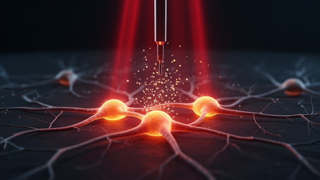
Dopamine, the neurotransmitter that governs movement, mood, cognition and reward, may one day be coaxed out of cells by nothing more than carefully engineered light. A team of researchers from Hefei University of Technology and several hospitals affiliated with Anhui Medical University, publishing in Frontiers in Photonics, reports that red and near-infrared light of specific wavelengths and doses can significantly stimulate the secretion and transformation of dopamine in retinal pigment epithelial cells. The finding, if it survives the journey from culture dish to clinic, could open a noninvasive route to treating myopia, Parkinson’s disease, Alzheimer’s disease, depression and schizophrenia, conditions in which dopaminergic signaling is either deficient or dysregulated.
The study rests on a deceptively simple observation that has long intrigued chronobiologists: dopamine activity in the body shows diurnal and seasonal variations, which the authors attribute to the influence of solar activity on the biological clock of the suprachiasmatic nucleus. Light exposure is known to stimulate dopamine biosynthesis in the retina by activating tyrosine hydroxylase, the rate-limiting enzyme that converts tyrosine into 3,4-dihydroxyphenylalanine, and by inducing aromatic L-amino acid decarboxylase, which catalyzes the formation of dopamine from levodopa. What remained unclear was whether light in the red and near-infrared band, the region most used in photobiomodulation, could drive dopamine secretion directly, and if so, which wavelengths and doses would be most effective.
To find out, the researchers irradiated ARPE-19 cells, a widely used model of the retinal pigment epithelium, with seven different light sources. Five were narrowband single-LED chips with emission peaks at 620, 680, 730, 800 and 850 nanometers. Two were phosphor-converted LEDs, built by coating mixtures of phosphors onto 450-nanometer blue-emitting chips, producing broadband spectra: model W710, with a peak at 710 nanometers spanning 600 to 850 nanometers, and model SW830, with a main peak at 830 nanometers and a secondary peak at 710 nanometers spanning 600 to 1,100 nanometers. Cells were exposed at a fixed power density of 3 milliwatts per square centimeter from a lamp height of 10 centimeters, with exposure times yielding energy densities of 1, 2, 4 and 8 joules per square centimeter. Control cells were handled identically but kept in the dark, and culture dishes were wrapped in tin foil to exclude stray light.
The choice of wavelengths was not arbitrary. In photobiomodulation, the dominant cellular mechanism involves cytochrome c oxidase, the terminal enzyme of the mitochondrial electron transport chain. The absorption peaks of its copper centers, reduced CuA, oxidized CuB, reduced CuB and oxidized CuA, sit at 620, 680, 760 and 825 nanometers respectively. The narrowband peaks selected at 620, 680, 730 and 850 nanometers align with or closely approach these absorption maxima, while the 800-nanometer peak falls between the reduced CuB and oxidized CuA bands. Crucially, the broadband SW830 source covers all four copper-center absorption peaks across its 600-to-1,100-nanometer span, and may additionally engage unknown mechanisms such as ion channels and nanostructured water layers in the cell membrane.
Dopamine concentrations in the cell supernatant were quantified by indirect enzyme-linked immunosorbent assay one hour after a second round of light treatment, with each test replicated twice. The results revealed a striking dependence on both wavelength and dose. Overall, dopamine levels in irradiated cells exceeded those of dark controls across all seven sources. Six of the seven sources, all except 850 nanometers, efficiently enhanced dopamine at 2 joules per square centimeter. The 850-nanometer source behaved anomalously, producing dopamine concentrations lower than every other wavelength at each corresponding energy level, with an abnormal dip at 2 joules and a partial recovery at 4 joules. The 680-nanometer source showed its own irregularity, with a dopamine concentration at 2 joules below that of other doses, a clue the authors interpret as possible activation of metabolic enzymes such as monoamine oxidase or catechol-O-methyltransferase that break dopamine down.
The broadband sources outperformed the narrowband chips in a critical respect: stability. Dopamine concentrations induced by W710 and SW830 were not only higher on average but remained relatively stable across the range of light doses, whereas narrowband-induced levels fluctuated considerably. Among all seven sources, SW830 was judged the best, delivering relatively high dopamine secretion with modest variation across doses. The authors attribute this to SW830’s full coverage of the cytochrome c oxidase absorption peaks and its possible stimulation of additional photophysiological pathways, suggesting that light-induced dopamine secretion involves the synergistic effect of multiple signals rather than a single molecular target.
Because dopamine synthesis draws on a chain of precursors, the team also tracked upstream and downstream metabolites by high-performance liquid chromatography. Glutamine can be hydrolyzed by glutaminase into glutamic acid, which serves as an indirect precursor; tyrosine is the direct substrate of tyrosine hydroxylase. Under irradiation with 680 nanometers and SW830, concentrations of glutamine, glutamic acid and tyrosine decreased rapidly with increasing light energy, consistent with their consumption in dopamine production. Under 850 nanometers, by contrast, these precursor concentrations barely moved, indicating that near-infrared light at this wavelength struggles to activate the synthetic reactions. Downstream, dopamine hydrochloride, norepinephrine hydrochloride and kynurenic acid rose sharply from 0 through 2 joules, peaked, and then declined slightly at higher doses under most conditions, explaining the drop in free dopamine at elevated doses as conversion into other compounds.
The interplay between dopamine and its metabolites proved more nuanced than a simple one-way synthesis. Dopamine, as an organic base, can neutralize with hydrochloric acid to form dopamine hydrochloride, which dissociates in the body into free dopamine and chloride ions, effectively storing the neurotransmitter for long-term function. The authors note that under SW830 irradiation, dopamine hydrochloride concentrations continued rising from 1 to 8 joules, which they argue explains how high free dopamine levels were sustained across the dose range. Dopamine also feeds the catecholamine pathway, serving as the precursor of norepinephrine and, further downstream, epinephrine. Meanwhile, kynurenic acid, a product of the tryptophan kynurenine pathway, competes with dopamine synthesis for shared cofactors such as tetrahydrobiopterin, linking light-induced dopamine dynamics to broader amino acid metabolism. Free dopamine itself is vulnerable to oxidation by high-energy photons, yet the low-energy photons of 850 or 830 nanometers cannot drive that reaction, which the authors suggest helps preserve the redox state of the neurotransmitter.
Cell morphology offered an independent readout of photophysiological effect. Under the microscope, ARPE-19 cells, typical adherent epithelial cells forming monolayers through tight junctions, showed polygonal atypia after light exposure. Cells cultured under 680 nanometers took on long-egg or slender-body shapes, while those under 850 nanometers and SW830 adopted round-egg or spheroid-body forms, with SW830 cells appearing rounder and more regular than either narrowband group. Cell density increased overall as exposure energy rose from 2 to 4 and then to 8 joules, with SW830 at 8 joules producing the largest density and 850 nanometers at 2 joules the smallest, reinforcing the impression that broadband near-infrared light is the gentlest and most productive of the tested conditions.
The work arrives amid growing evidence that light and dopamine are deeply intertwined. Prior studies have shown that delivering 710-nanometer far-red light into the substantia nigra pars compacta of mice via micro-optical fiber enhanced dopamine neuron firing four-fold, from 2.2 to 8.8 impulses per second, while visible fluorescent light has been reported to harm dopamine neurons and induce neurodegeneration. Deep-brain light stimulation at 840 nanometers in rats has been shown to decrease glutamate while increasing dopamine in the subthalamic nucleus, hinting at therapeutic potential for Parkinson’s disease. The new study extends these findings by proposing a noninvasive alternative: radiating tissue from outside the body with broadband near-infrared light. The authors acknowledge the limitations of their in vitro approach, noting that animal or human models with dopamine measured in blood or cerebrospinal fluid by liquid chromatography would yield more accurate values, and they indicate that further work on Parkinson’s disease model mice is forthcoming. If those experiments bear out the cell-culture results, engineered light could become a prescription for the brain’s most celebrated messenger.
Subject of Research: science news
Article Title: Light-induced secretion and transformation of neurotransmitter dopamine
Article References: Wei, S., Liu, C., Wu, Z., Si, J., Huo, X., Shen, C., Zhen, Y., Liu, D., & Chen, L. (2025). Light-induced secretion and transformation of neurotransmitter dopamine. Frontiers in Photonics, 6, Article 1647467. https://doi.org/10.3389/fphot.2025.1647467
Image Credits: AI Generated
DOI: 10.3389/fphot.2025.1647467
Keywords: Light-induced, secretion, transformation, neurotransmitter, dopamine, scientific research
Cite Scienmag News
Diana Fleming. (October 17, 2026). Red and Near-Infrared Light Can Trigger Dopamine Release in Cells. Scienmag. https://scienmag.com/red-and-near-infrared-light-can-trigger-dopamine-release-in-cells/
Diana Fleming. "Red and Near-Infrared Light Can Trigger Dopamine Release in Cells." Scienmag, 17 October 2026, https://scienmag.com/red-and-near-infrared-light-can-trigger-dopamine-release-in-cells/. Accessed 17 October 2026.
Diana Fleming. "Red and Near-Infrared Light Can Trigger Dopamine Release in Cells." Scienmag. October 17, 2026. https://scienmag.com/red-and-near-infrared-light-can-trigger-dopamine-release-in-cells/


