


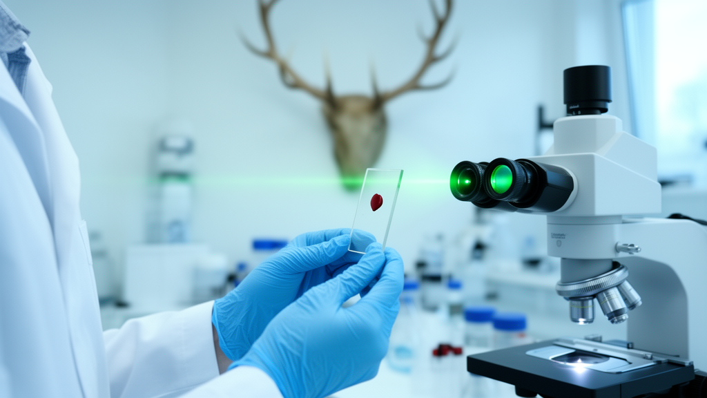
Determining the sex of a wild animal usually requires something investigators rarely have: the animal itself, intact and identifiable. Once only blood remains — collected during a brief sedation, recovered from a decomposed carcass, or preserved in a freezer years before anyone thought to record whether the donor was male or female — most forensic options fall away. A new study from Texas Tech University, published in Frontiers in Analytical Science, shows that a beam of laser light and a machine-learning algorithm may be enough to answer the question. Working with white-tailed deer (Odocoileus virginianus), the team demonstrated that Raman spectroscopy combined with Partial Least Squares Discriminant Analysis (PLS-DA) can classify the sex of a deer from serum or plasma with accuracies that, under favorable conditions, exceed 98 percent.
The technique exploits a fundamental property of molecules. When monochromatic laser light strikes a sample, most photons scatter elastically, keeping their energy unchanged. But a small fraction exchange energy with molecular vibrations, emerging shifted in wavelength in ways that are unique to the chemical bonds they encountered. This inelastic scattering produces the Raman spectrum: a fingerprint of peaks corresponding to proteins, lipids, nucleic acids, and carbohydrates in the sample. Because sex hormones such as testosterone and estrogen reshape metabolism — altering lipid content, protein profiles, immune function, and stress responses — the biochemical state of a male and a female animal differs measurably, and those differences leave detectable traces in the vibrational spectra of blood-derived fluids. Crucially, the method is label-free, requires minimal sample preparation, and does not destroy the specimen.
The research team, led by Md Salahuddin Majumder with Ernest Smith and Lenka Halámková, worked with ten captive white-tailed deer, five of each sex, aged one to six years, under an approved animal care protocol. Blood was collected aseptically, processed into serum and plasma, and stored at −80 °C before analysis. Using a ThermoFisher DXR3 Raman microscope with a 785-nanometer excitation laser set at 30 milliwatts, they acquired spectra from dried droplets on foil-covered slides, sampling three non-overlapping spots per droplet with three replicate scans each. The careful sampling scheme yielded 36 serum spectra and 36 plasma spectra per animal — 360 spectra for each biofluid, and a total of 720 spectra feeding into the classification models. Spectral resolution was set at 2 cm⁻¹ across a range from 399 to 3,299 cm⁻¹.
Raw Raman spectra from biological fluids are notoriously messy, dominated by fluorescence background and inter-sample variability, so preprocessing mattered. The team applied automatic weighted least squares baseline correction, 1-norm area normalization, and mean-centering before importing the data into MATLAB with the PLS Toolbox. PLS-DA, a supervised multivariate method, then compressed the thousands of correlated wavenumber variables into a small set of latent variables chosen to maximize separation between the male and female classes. Model quality was assessed through classification error rates, Matthews Correlation Coefficient (MCC), prediction R², and root mean square error, with receiver operating characteristic (ROC) analysis providing a threshold-independent measure of discrimination.
Because only ten animals were available, validation design was central to the study’s credibility. The researchers systematically tested four calibration-to-validation splits — 1:4, 2:3, 3:2, and 4:1 — always partitioning by whole animal so that no individual contributed spectra to both training and testing sets. The serum-based models delivered striking results. With severely limited training data (just two deer), accuracy dropped to 79.2 percent. But once calibration was more balanced, performance soared: 99.1 percent for the 2:3 split, 99.3 percent for the 3:2 split, and 98.6 percent for the 4:1 split, with MCC values approaching 1.00 and ROC areas under the curve of 0.998 to 1.00. The full model, trained on all 360 serum spectra, reached 99.4 percent accuracy with an error rate of just 0.56 percent.
Plasma told a more complicated story. Although the full plasma model achieved 99.2 percent internal accuracy, external validation was erratic: 85.8, 82.4, and 86.1 percent in the first three splits, and a startling collapse to 57.0 percent in the 4:1 configuration — barely better than a coin flip. The authors attribute this not necessarily to a worse model but to the very small validation set of only two animals, combined with the greater biochemical variability of plasma. Plasma retains fibrinogen, clotting factors, and anticoagulant effects that serum lacks, introducing spectral noise that makes sex-specific signatures harder to isolate. Serum preparation removes these unstable components, producing cleaner, more reproducible profiles — and the data bore this out, with serum consistently outperforming plasma across every metric.
The spectral differences underpinning the classification were subtle but biochemically coherent. Male deer showed enhanced lipid-associated signals at approximately 1,315–1,330 cm⁻¹ (CH₂ twisting) and 1,445 cm⁻¹ (CH₂/CH₃ bending), while female deer displayed stronger protein-related peaks in the amide III region (1,220–1,280 cm⁻¹) and the amide I/II region (1,540–1,630 cm⁻¹). Notably, these regions overlap with known Raman bands of sex hormones — testosterone signals near 786, 856, 1,490, and 1,636 cm⁻¹, and female hormones in the 682–902 and 1,291–1,625 cm⁻¹ ranges — suggesting the hormonal differences between sexes directly shape the spectral fingerprints. Intriguingly, male samples clustered more tightly in the latent space while female samples dispersed more widely, a pattern the authors link to hormonal fluctuations and reproductive variability, and one consistent with prior Raman studies of saliva and bloodstains.
To confirm the finding was not an artifact of one algorithm, the team repeated the comparison using a second approach: principal component analysis followed by linear discriminant analysis (PCA-LDA), a standard two-step method in vibrational spectroscopy. The results were reassuringly convergent. For serum, PLS-DA achieved 99.1 percent external accuracy while PCA-LDA reached 92.6 percent; for plasma, the two methods landed within a fraction of a percent of each other (82.4 versus 82.9 percent). The method-independent success of serum classification strengthens the argument that sex-specific spectral signatures are genuine biological signals rather than statistical artifacts of a particular model.
The implications reach well beyond deer. In poaching cases, wildlife trafficking investigations, and enforcement of sex-specific hunting regulations, sex can determine whether a crime has occurred at all. For conservation biologists, sex ratios are core demographic parameters guiding population models, breeding programs, and protection laws. Current genetic methods — PCR amplification of sex chromosome-linked genes such as SRY or Amelogenin — are accurate but demand specialized laboratories and intact DNA, which degrades quickly in non-invasive or archived samples. Raman spectroscopy, by contrast, tolerates degraded material, requires no reagents, and is increasingly available in portable formats. The authors envision investigators analyzing dried blood spots in the field, with portable instruments delivering near-immediate results — a capability that could prove decisive for species regulated under CITES, where sex-specific rules apply.
Caveats remain, and the researchers are candid about them. Ten animals cannot represent the full physiological diversity of a species whose hormonal cycles, age structure, and seasonal reproduction all modulate blood chemistry, and future validation across larger cohorts, populations, and reproductive states is essential before courtroom deployment. Seasonal sampling of females, in particular, may influence spectral dispersion and classification accuracy. Still, as a proof of concept, the study marks a genuine milestone: the first demonstration that blood-derived fluids alone, interrogated only by light and statistics, can reveal the sex of one of North America’s most ecologically and economically important cervids. If portable Raman systems and automated analysis pipelines mature as expected, the humble drop of blood may become a forensic witness in its own right.
Subject of Research: Sex determination of white-tailed deer from blood-derived biofluids using Raman spectroscopy and PLS-DA chemometric classification
Article Title: Sex determination of white-tailed-deer (Odocoileus virginianus) from plasma and serum samples by using Raman spectroscopy and PLS-DA method: a forensic perspective
Article References: Majumder, M. S., Smith, E., & Halámková, L. (2026). Sex determination of white-tailed-deer (Odocoileus virginianus) from plasma and serum samples by using Raman spectroscopy and PLS-DA method: a forensic perspective. Frontiers in Analytical Science, 5, Article 1727520. https://doi.org/10.3389/frans.2025.1727520
Image Credits: AI Generated
DOI: 10.3389/frans.2025.1727520
Keywords: Raman spectroscopy, PLS-DA, white-tailed deer, wildlife forensics, sex determination, serum, plasma, chemometrics, machine learning, vibrational spectroscopy, conservation, Odocoileus virginianus
Cite Scienmag News
Margaret Porter. (October 16, 2026). Raman Spectroscopy Can Tell Male Deer From Female Using a Drop of Blood. Scienmag. https://scienmag.com/raman-spectroscopy-can-tell-male-deer-from-female-using-a-drop-of-blood/
Margaret Porter. "Raman Spectroscopy Can Tell Male Deer From Female Using a Drop of Blood." Scienmag, 16 October 2026, https://scienmag.com/raman-spectroscopy-can-tell-male-deer-from-female-using-a-drop-of-blood/. Accessed 16 October 2026.
Margaret Porter. "Raman Spectroscopy Can Tell Male Deer From Female Using a Drop of Blood." Scienmag. October 16, 2026. https://scienmag.com/raman-spectroscopy-can-tell-male-deer-from-female-using-a-drop-of-blood/


