


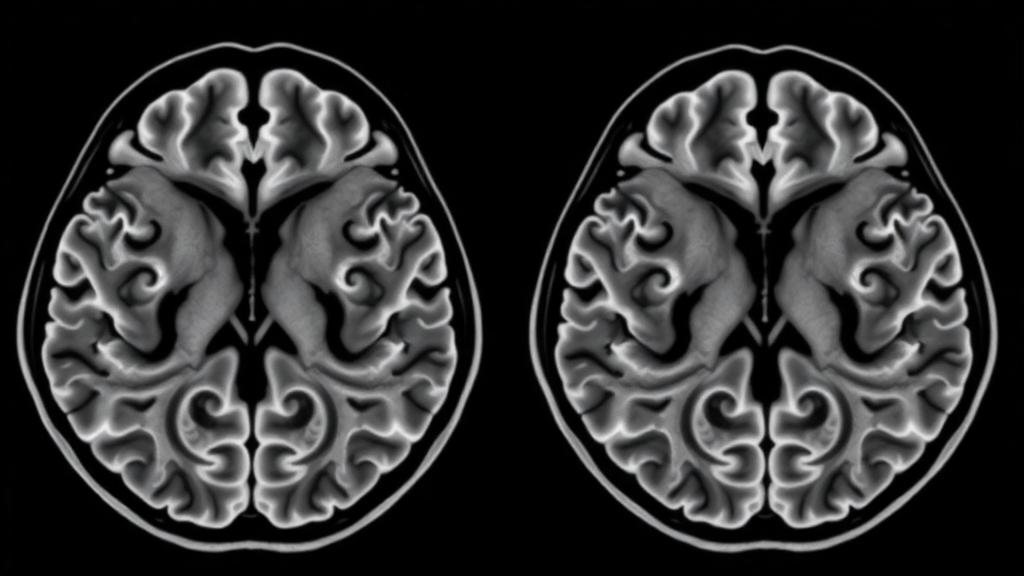
Polymicrogyria has long been one of the most confounding diagnoses in pediatric neurology, a condition in which the surface of the brain develops far too many small, abnormally folded ridges. Clinicians have traditionally encountered it as a finished product, visible on scans of children who present with epilepsy, developmental delay, or muscle tone abnormalities. But a new study published in Pediatric Radiology has done something unusually rare: it captured the malformation at the very moment of its emergence, in fetuses of just 21 weeks’ gestational age, by combining high-resolution post-mortem magnetic resonance imaging with detailed neuropathological examination. The result is a kind of time-lapse photograph of a brain folding wrongly, and it offers some of the clearest evidence yet that polymicrogyria is not one disease but several distinct processes converging on the same devastating endpoint.
The research team, led by Giana Izzo and Andrea Righini of Ospedale dei Bambini Vittore Buzzi in Milan together with colleagues at the University of Naples Federico II and the University of Milan, studied four fetuses in which fetal MRI had raised suspicion of a cortical malformation. After termination of pregnancy following multidisciplinary counseling, each fetus underwent post-mortem MRI within 24 hours of delivery, on fresh specimens stored at 4 to 5 degrees Celsius, followed by complete neuropathological workup including immunohistochemistry for markers such as NeuN, GFAP, CD34, CD3, CD8, and myelin basic protein. The four cases were deliberately chosen to represent the major etiological categories of polymicrogyria: an ischemic form, an infectious form caused by congenital cytomegalovirus, and two genetic forms driven by mutations in PIK3CA and ADGRG1/GPR56.
What makes the study technically remarkable is the imaging resolution achieved. The post-mortem examinations were performed on a 1.5-Tesla scanner using high-resolution T2-weighted fast spin-echo sequences with an in-plane resolution of roughly 0.26 square millimeters, approximately four times finer than what in vivo fetal MRI can achieve. At 21 weeks of gestation, the fetal cortex is organized into a transient laminar architecture, with distinct layers including the cortical plate, the subplate containing thalamocortical fibers, the intermediate zone, the periventricular zone, and the germinal zone. Post-mortem MRI could resolve these layers individually, allowing the researchers to detect precisely where and how the normal laminar pattern broke down in each case, something in vivo fetal MRI, limited by motion artifacts and field strength, could only hint at as vague cortical irregularity.
The first case illustrated the ischemic pathway. This fetus was the surviving co-twin of a monochorionic pregnancy complicated by twin-to-twin transfusion syndrome treated with laser therapy, after which the donor twin died in utero. Fetal MRI performed roughly two weeks after the procedure, following a prior scan that had shown no abnormalities, revealed volume loss in one hemisphere and a wart-like irregularity of the cortical plate in perisylvian frontoparietal regions. Post-mortem MRI sharpened the picture considerably, showing a wavy, irregularly thickened cortical plate that appeared to extend beyond its expected boundary, a markedly reduced subplate, and an irregular thickening of the leptomeningeal layer. Histology confirmed the classic festooned appearance of polymicrogyria and revealed foci of laminar necrosis, micro-regional disruption of the pial basement membrane, ectopic neurons and glial cells outside the cortex, macrophage infiltration, and proliferating meningeal vessels, all signatures of an acute, regionally selective vascular insult striking a developing brain.
The second case, driven by congenital cytomegalovirus infection confirmed by PCR of amniotic fluid, presented a fundamentally different pattern. CMV has a well-documented tropism for radial glial progenitor cells, and the imaging showed bilateral but strikingly asynchronous injury. One hemisphere already displayed an established polymicrogyria pattern, with a thickened irregular cortical plate, an indistinct subplate, a disrupted germinal zone, and a markedly hypointense rim along the cortical ribbon on T2-weighted images, corresponding to laminar necrosis and calcifications that matched areas of pronounced diffusion restriction seen on in vivo imaging. The other hemisphere showed an earlier stage of the same process, with edematous T2 hyperintensity, a thickened cortical plate poorly delineated from the subplate, and, notably, an undulating appearance of the thalamocortical fiber band, as if the cortical plate were crumpling onto the subplate beneath it. Immunohistochemistry confirmed diffuse laminar disorganization, intense reactive gliosis, lympho-macrophage infiltration on CD3 and CD8 staining, and, crucially, an intact pial basement membrane, distinguishing this inflammatory mechanism from the ischemic case.
The two genetic cases demonstrated that malformations arising from mutations can look entirely different from acquired forms. The third fetus carried a somatic mosaic PIK3CA mutation, identified through next-generation sequencing of a biopsy from an overgrown limb, and showed hemimegalencephaly with unilateral polymicrogyria. Post-mortem MRI revealed expansion and blurring of the intermediate zone and subplate, globally reduced T2 signal suggesting increased cellularity in every layer, and loss of the normal demarcation of thalamocortical fibers. Histology found an excess of mature neurons in the superficial cortical layers, an inversion of the normal inside-out neuronal layering, while the pial basement membrane and leptomeninges remained intact and no gliosis was present. The researchers attribute this profile to hyperactivation of the PI3K-AKT signaling pathway, driving progenitor hyperproliferation and impaired apoptosis rather than tissue destruction.
The fourth case involved a mutation in ADGRG1, also known as GPR56, a gene encoding a receptor expressed on the end feet of radial glial cells that anchors them to the pial basement membrane. When that anchorage fails, neurons overmigrate through gaps in the pial barrier, producing a mixed picture. Fetal MRI had shown frontal bumps along the cortical rim, subependymal heterotopic nodules, cerebellar vermian hypoplasia, and flattening of the pons. Post-mortem MRI delineated a thickened, irregular cortical plate with focal mushroom-like protrusions and a cobblestone-like surface at the frontal vertex, alongside polymicrogyria-like changes in parietal regions. Histology confirmed the coexistence of both patterns: festooning cortical bands with vessels entrapped in fused sulci characteristic of polymicrogyria, and complete laminar disorganization with meningeal vessels trapped in the subplate, pial basement membrane discontinuity, and neuronal overmigration characteristic of cobblestone cortex, placing this condition within the mechanistic spectrum of dystroglycanopathies.
Taken together, the four cases allow the authors to propose a set of differentiating imaging features that map onto underlying mechanisms. Symmetry of involvement separates the bilateral, asynchronous infectious form from the vascular-territory-limited ischemic form and the hemispheric overgrowth of PIK3CA disease. The integrity of the pial basement membrane divides the cases sharply: it is focally breached in ischemic injury and diffusely disrupted in ADGRG1-related disease, but preserved in both CMV infection and PIK3CA-related overgrowth. The presence of an extracortical layer, the pattern of laminar alteration, the state of the subplate, and associated anomalies such as cerebellar hypoplasia or hemispheric enlargement each carry diagnostic weight. In the CMV case, the undulating thalamocortical fiber band stands out as a particularly evocative early marker, suggesting that disturbance of the cortical plate-subplate interface may be a harbinger of abnormal folding before any overt gyral anomaly appears.
The authors are careful about the limits of their work. The series comprises only four deliberately selected fetuses, examined retrospectively and without blinded image interpretation, and post-mortem correlation was available only in cases of pregnancy termination, which may not represent the full clinical spectrum of polymicrogyria. No quantitative measurements of cortical thickness were performed, the fetal imaging was acquired at 1.5 Tesla rather than the higher-resolution 3-Tesla systems now optimized for fetal work, and the interval between prenatal and post-mortem scanning may have introduced tissue changes affecting comparison. Direct translation of post-mortem findings to routine in vivo fetal MRI remains constrained by differences in acquisition conditions and tissue characteristics. The patterns described are therefore framed as hypothesis-generating observations that require validation in larger, independent cohorts with standardized quantitative approaches.
Even with those caveats, the conceptual payoff is substantial. The study reinforces the view, increasingly dominant in developmental neuropathology, that polymicrogyria is a final common pathway rather than a single entity, shaped by genetic mutations, vascular insults, viral infection, and metabolic disturbance acting at different moments of corticogenesis. It also elevates the pial surface and its basement membrane to a central orchestrating role in normal cortical development, since the integrity or breakdown of that interface emerges as the key anatomical hinge separating the major pathogenic routes. For families facing a prenatal diagnosis of a cortical malformation, the practical hope is that recognizing these early signatures, particularly the state of the laminar pattern and the pial boundary, will sharpen differential diagnosis, guide targeted genetic testing, and ultimately improve the counseling offered for future pregnancies, turning a descriptive label into a mechanistic explanation.
Subject of Research: Early laminar disruption in fetal incipient polymicrogyria characterized by post-mortem MRI and neuropathological correlation
Article Title: Laminar pattern disruption in fetal incipient polymicrogyria: insights from post-mortem magnetic resonance imaging and neuropathological correlation
Article References: Izzo, G., Tortora, M., Toto, V., Lanna, M., Doneda, C., Arrigoni, F., Tortora, F., Parazzini, C., & Righini, A. (2026). Laminar pattern disruption in fetal incipient polymicrogyria: insights from post-mortem magnetic resonance imaging and neuropathological correlation. Pediatric Radiology. https://doi.org/10.1007/s00247-026-06770-4
Image Credits: AI Generated
DOI: 10.1007/s00247-026-06770-4
Keywords: polymicrogyria, fetal MRI, post-mortem MRI, cortical development, neuropathology, cytomegalovirus, PIK3CA, ADGRG1, pial basement membrane, cortical lamination, twin-to-twin transfusion syndrome, Pediatric Radiology
Cite Scienmag News
Colin Clarke. (October 6, 2026). Post-mortem MRI reveals how fetal brains first begin to form the folds of polymicrogyria. Scienmag. https://scienmag.com/post-mortem-mri-reveals-how-fetal-brains-first-begin-to-form-the-folds-of-polymicrogyria/
Colin Clarke. "Post-mortem MRI reveals how fetal brains first begin to form the folds of polymicrogyria." Scienmag, 6 October 2026, https://scienmag.com/post-mortem-mri-reveals-how-fetal-brains-first-begin-to-form-the-folds-of-polymicrogyria/. Accessed 6 October 2026.
Colin Clarke. "Post-mortem MRI reveals how fetal brains first begin to form the folds of polymicrogyria." Scienmag. October 6, 2026. https://scienmag.com/post-mortem-mri-reveals-how-fetal-brains-first-begin-to-form-the-folds-of-polymicrogyria/


