


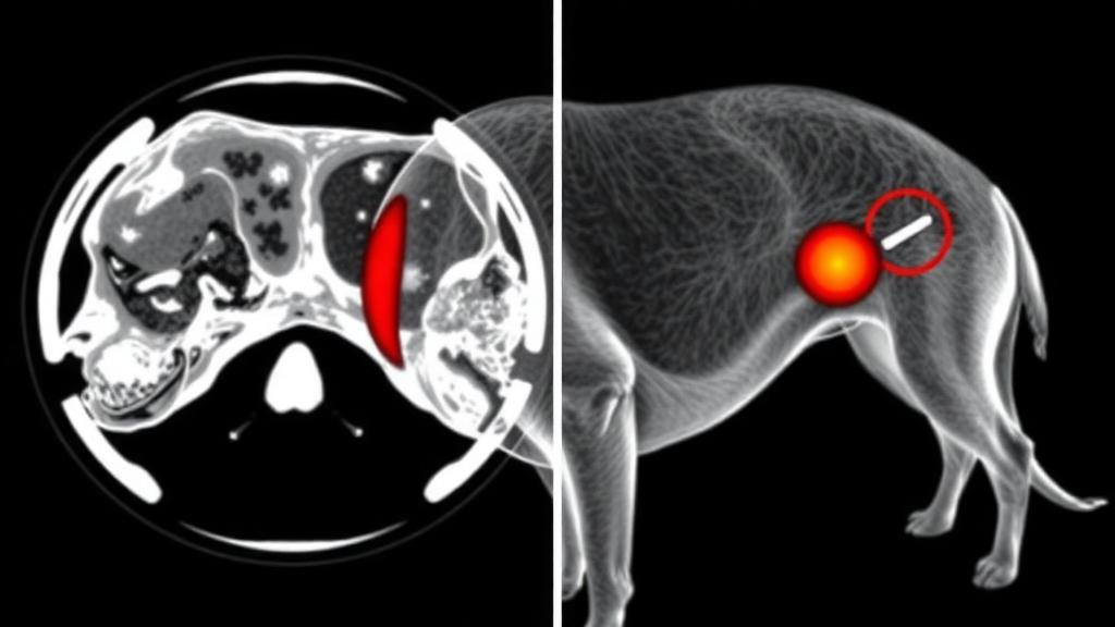
In a first for veterinary oncology, researchers have documented the detailed positron emission tomography findings and standardized uptake values in a dog with apocrine gland anal sac adenocarcinoma, one of the most quietly aggressive cancers in companion animals. The case, reported by a surgical team at Chungbuk National University in South Korea and published in Frontiers in Veterinary Science, describes how a hybrid imaging technique borrowed from human medicine mapped the true extent of a small dog’s cancer before a single incision was made, and how those metabolic fingerprints guided a surgery that reached deeper into the abdomen than conventional imaging alone would have suggested.
Apocrine gland anal sac adenocarcinoma, abbreviated AGASACA, is a malignant epithelial tumor arising from the anal sacs, the small scent glands tucked on either side of a dog’s anus. It is not common, accounting for roughly 2 percent of all skin tumors and about 17 percent of perianal malignancies in dogs, but it is notorious for two reasons. First, it frequently spreads to regional lymph nodes, sometimes even when the primary tumor is small. Second, it often triggers a paraneoplastic syndrome in which the tumor produces hormone-like signals that drive blood calcium to dangerously high levels, causing increased thirst, weakness, and strain on the kidneys. Dogs typically arrive at the clinic with constipation or straining to defecate, caused by the primary mass or by enlarged metastatic lymph nodes pressing on the rectum.
The patient in this report was a 10-year-old castrated male Maltese weighing just 3.88 kilograms, referred for evaluation of a right anal sac mass and enlarged intra-abdominal lymph nodes. Fine-needle aspiration performed at the referring clinic had already revealed cells with cytological features of malignancy, but a definitive diagnosis would have to wait for histopathology after surgery. The clinical picture was telling: the dog had mild leukocytosis, an elevated ionized calcium concentration of 1.66 mmol/L against a reference interval of 1.16 to 1.40 mmol/L, and metabolic acidosis marked by a raised anion gap, low bicarbonate, and negative base excess. Chest radiographs showed no evidence of pulmonary metastasis, but the abdominal picture remained uncertain.
That uncertainty is exactly where 18F-fluorodeoxyglucose positron emission tomography combined with computed tomography, or 18F-FDG PET/CT, enters the story. The technique exploits a fundamental quirk of cancer biology: malignant cells burn glucose at an accelerated rate. The radiopharmaceutical 18F-FDG is a glucose analog tagged with a positron-emitting isotope of fluorine. Once injected, it is taken up by metabolically hungry tissues, where it becomes trapped after phosphorylation, allowing the PET scanner to map glucose consumption across the entire body. Fused with the anatomical detail of CT, the result is an image that shows not just where structures are, but which of them are biologically active. Uptake is quantified with the standardized uptake value, calculated by dividing the tissue concentration of FDG in megabecquerels per milliliter by the injected activity per gram of body weight. The maximum value within a region of interest, SUVmax, serves as a compact summary of a lesion’s metabolic intensity.
In human oncology, FDG PET/CT is an established staging workhorse, outperforming CT alone for assessing disease extent across many tumor types. In veterinary medicine it has been explored in canine lymphoma, osteosarcoma, mammary carcinoma, cutaneous squamous cell carcinoma, and granulosa cell tumors, but its role in specific tumor entities remains poorly defined. For AGASACA, PET/CT had been mentioned as a staging option, yet no detailed imaging findings or SUV measurements had ever been published. The Korean team set out to fill that gap with a rigorously documented single case.
The imaging protocol was meticulous. The dog was fasted for at least 12 hours, anesthetized with medetomidine and propofol, intubated, and maintained on isoflurane in 100 percent oxygen with mechanical ventilation and intravenous fluids at twice the maintenance rate to support blood pressure. After anesthetic induction, 0.187 mCi/kg of 18F-FDG was injected intravenously. Whole-body CT was acquired on a multidetector scanner at 100 mAs and 120 kVp with 1.25 mm slice thickness, before and after contrast enhancement with iohexol. PET followed on the same scanner after a 60-minute uptake period, with the entire imaging session lasting about two hours. Regions of interest were manually delineated on the fused images, and SUVmax was measured for each suspicious lesion.
The results were striking. CT identified a mass measuring 3.8 by 2.6 by 3.8 centimeters within the right anal sac, an internal iliac lymph node of 3.5 by 2.2 by 1.6 centimeters, and a lumbar aortic lymph node of 1.3 by 1.0 by 1.3 centimeters. PET revealed focally intense FDG uptake in all three: SUVmax 7.3 in the primary tumor, 6.35 in the internal iliac node, and 4.3 in the lumbar aortic node. Crucially, no distant metastatic uptake appeared in the lungs, liver, spleen, or other visceral organs. The dog was classified as Stage IIIa, defined as locoregional nodal metastasis with a maximum metastatic node smaller than 4.5 centimeters and no distant spread. The nodal SUVmax values clearly exceeded the general threshold range of 2.5 to 3.5 used to flag metabolically active lymph nodes as suspicious for metastasis.
Guided by the metabolic map, the surgeons performed an intra-abdominal lymphadenectomy before the right anal sacculectomy, sequencing the procedure to minimize contamination of the abdominal field. The enlarged lumbar aortic node, found adjacent to the aorta at the level of the caudal pole of the right kidney, consisted of two distinct segments that were harvested separately. The enlarged internal iliac node was then excised. During removal of the anal sac, gross invasion of part of the rectal wall was discovered, and the involved portion was resected along with the primary mass. Histopathology confirmed AGASACA in the primary tumor, showing tubular to acinar differentiation, moderate anisocytosis and anisokaryosis, necrosis, and desmoplasia, with a mitotic count of 30 per 2.37 square millimeters and no vascular invasion. The resected lymph nodes contained morphologically similar metastatic adenocarcinoma, with mitotic counts of 35 and 30 in the two lumbar aortic segments and 16 in the internal iliac node, confirming the preoperative Stage IIIa classification.
The postoperative course offered both encouragement and a sobering lesson. The day after surgery, ionized calcium fell to 1.35 mmol/L, back within the reference interval, demonstrating that the paraneoplastic hypercalcemia resolved with removal of the tumor burden. A surgical site dehiscence required a secondary repair, and the dog was discharged 12 days after the initial operation, initially doing well with a good appetite and no fecal incontinence. But at approximately three months, thoracic radiography and abdominal ultrasonography revealed enlargement of multiple intra-abdominal lymph nodes, the rectum was displaced ventrally by a nodal mass, and hypercalcemia recurred at 2.31 mmol/L, signaling progressive nodal metastasis. The owner declined further tumor-directed treatment, and the dog was euthanized on postoperative day 104.
The discussion surrounding the case highlights why the findings matter. Conventional staging for AGASACA relies on abdominal ultrasonography and CT, which judge lymph nodes by size and morphology and say nothing about metabolic activity. Fine-needle aspiration can confirm cytology but is invasive and technically difficult for deep nodes. Lymphatic drainage in AGASACA is also unpredictable: in a prospective pilot study of eight dogs with unilateral tumors, preoperative and intraoperative sentinel lymph node mapping disagreed in four dogs, and contralateral sentinel nodes were identified in four of eight cases. Lumbar aortic involvement, seen here, is less commonly reported than disease in the iliosacral lymphocenter. The primary tumor’s SUVmax of 7.3 sits within the range reported for other canine carcinomas, including adenocarcinomas at 7.6 to 27.0 and malignant mammary carcinomas at 2.97 to 10.01, though cross-study comparisons remain hazardous because scanner hardware and reconstruction algorithms vary. Intriguingly, the internal iliac node showed higher uptake despite a lower mitotic count than the lumbar aortic node, a reminder that FDG uptake reflects cellular composition and glucose metabolism, not proliferation alone. As a single case, the values cannot be generalized, and no disease-specific reference range yet exists for canine AGASACA. But the authors argue the case demonstrates that PET/CT can complement morphology-based imaging by exposing metabolically active nodes at both typical and atypical sites, potentially sharpening preoperative staging and surgical planning. For a cancer that so often hides its true spread until the abdomen is open, that metabolic window may prove to be a genuinely valuable addition to the veterinary oncologist’s toolkit.
Subject of Research: First reported 18F-FDG PET/CT imaging findings and SUVmax values in a dog with apocrine gland anal sac adenocarcinoma and confirmed lymph node metastases
Article Title: Case Report: 18F-FDG PET/CT findings and SUVmax characterization in a dog with apocrine gland anal sac adenocarcinoma
Article References: Shim, B., Kim, Y., & Lee, S. (2026). Case Report: 18F-FDG PET/CT findings and SUVmax characterization in a dog with apocrine gland anal sac adenocarcinoma. Frontiers in Veterinary Science, 13, Article 1977879. https://doi.org/10.3389/fvets.2026.1977879
Image Credits: AI Generated
DOI: 10.3389/fvets.2026.1977879
Keywords: 18F-FDG PET/CT, apocrine gland anal sac adenocarcinoma, SUVmax, veterinary oncology, canine cancer, lymph node metastasis, staging, paraneoplastic hypercalcemia, nuclear medicine, case report, comparative oncology, anal sacculectomy
Cite Scienmag News
Nathaniel Bowman. (October 16, 2026). PET Scan Reveals Hidden Cancer Spread in a Dog With Anal Sac Tumor. Scienmag. https://scienmag.com/pet-scan-reveals-hidden-cancer-spread-in-a-dog-with-anal-sac-tumor/
Nathaniel Bowman. "PET Scan Reveals Hidden Cancer Spread in a Dog With Anal Sac Tumor." Scienmag, 16 October 2026, https://scienmag.com/pet-scan-reveals-hidden-cancer-spread-in-a-dog-with-anal-sac-tumor/. Accessed 16 October 2026.
Nathaniel Bowman. "PET Scan Reveals Hidden Cancer Spread in a Dog With Anal Sac Tumor." Scienmag. October 16, 2026. https://scienmag.com/pet-scan-reveals-hidden-cancer-spread-in-a-dog-with-anal-sac-tumor/


