


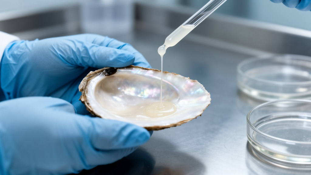
The road to younger-looking skin may run through one of the ocean’s most unglamorous secretions. In a study published in Frontiers in Natural Products, researchers report that a simple water extract of mucus from the Akoya pearl oyster, Pinctada fucata martensii, protected mouse skin and cultured cells from ultraviolet A radiation, the wavelength band most responsible for deep-tissue photoaging. The extract, designated AKN, was prepared without enzymes, solvents, or harsh chemistry, and its protective performance in animals matched that of ergothioneine, a well-studied antioxidant used as a benchmark in photoprotection research.
The significance of the work lies partly in what it did not do. Most previous investigations of bioactive compounds from P. martensii, a commercially vital aquaculture species farmed along the coasts of southern China and southwestern Japan, have relied on enzymatic hydrolysis, in which trypsin, alcalase, or flavourzyme chop discarded oyster flesh into short peptides with antioxidant or anti-inflammatory activity. Enzymatic digestion substantially reshapes the native molecular profile and risks destroying sensitive bioactive sequences. The new study instead collected roughly one liter of fresh mucus from 100 oysters during routine shucking, diluted it with ultrapure water, homogenized it at 4 degrees Celsius, centrifuged the mixture, filtered it through 0.45 and 0.22 micrometer membranes, and passed the filtrate through a 3 kilodalton molecular-weight-cutoff membrane. The low-molecular-weight permeate was then freeze-dried. This hydrolysis-free workflow preserves the mucus’s native cocktail of free amino acids, small peptides, nucleosides, and polar metabolites exactly as the oyster secretes them.
Chemical profiling revealed a composition dominated by small, water-soluble osmolytes. Targeted quantification showed that taurine accounted for 8.0 percent of the dry weight and betaine for 4.1 percent, while total amino acids measured after acid hydrolysis reached 6.0 percent. Inorganic components, chiefly chloride and sodium followed by sulfate, bicarbonate, potassium, and magnesium, made up 72.0 percent of the dry mass, and total sugars were minimal at 0.15 percent. Glycine was the predominant amino acid, followed by glutamic acid, proline, alanine, and valine. Untargeted liquid chromatography-mass spectrometry, performed under two orthogonal separation modes, reversed-phase C18 and hydrophilic interaction chromatography, generated distinct chemical fingerprints in which taurine and betaine appeared prominently under both conditions, and a feature putatively assigned to hypoxanthine emerged only under the HILIC mode. Because authentic standards were not run under identical chromatographic conditions, the authors treated these annotations as putative, a methodological caution that underscores how much of the extract’s chemistry remains uncharacterized.
With the extract characterized, the team turned to cellular models. In L929 murine fibroblasts, the workhorse cell type of connective tissue research, a single UVA dose of 10 joules per square centimeter, delivered at a peak wavelength of 365 nanometers, drove the cells into a senescent state. Prior toxicity testing across concentrations from 7.81 to 1,000 micrograms per milliliter had confirmed that AKN alone was non-cytotoxic, maintaining viability above 95 percent. When AKN was administered 12 hours before irradiation at 400 micrograms per milliliter, it significantly suppressed the mRNA levels of p16, p21, and p53, the canonical cell-cycle arrest genes whose upregulation marks premature cellular senescence. Senescence-associated beta-galactosidase staining, the gold-standard histochemical marker of the senescent phenotype, corroborated the molecular data: UVA-irradiated fibroblasts displayed the characteristic enlarged, flattened morphology and a pronounced increase in blue-stained cells, while AKN treatment markedly reduced the proportion of senescence-positive cells and largely restored normal morphology.
The extract also showed anti-inflammatory capacity in a second cellular model. BV2 murine microglial cells stimulated with lipopolysaccharide, a bacterial endotoxin that activates toll-like receptor 4-dependent inflammatory signaling, responded to AKN co-treatment with significantly reduced mRNA expression of the pro-inflammatory cytokines TNF-alpha, IL-6, and IL-1beta. AKN simultaneously lowered intracellular reactive oxygen species, measured by DCFH-DA fluorescence. This dual action on oxidative and inflammatory axes is mechanistically coherent with current models of photoaging, in which UVA-generated reactive oxygen species overwhelm endogenous antioxidant defenses, activate the nuclear factor-kappaB axis, and trigger a self-reinforcing feed-forward cascade linking oxidative stress, inflammatory signaling, the senescence-associated secretory phenotype, and irreversible cell-cycle arrest.
The decisive test came in living animals. Six- to seven-week-old male BALB/c mice were randomly assigned to five groups of six animals each: an untreated control, a UVA model group, and three treatment groups receiving topical applications of 1 milligram per milliliter AKN, 4 milligrams per milliliter AKN, or 4 milligrams per milliliter ergothioneine. Thirty minutes before each irradiation session, 200 microliters of the corresponding solution were applied to shaved dorsal skin, and the mice were exposed to UVA at 400 millijoules per square centimeter per session for 16 days, with investigators blinded to treatment allocation. By the end of the experiment, the UVA model group had developed the classic macroscopic photoaging phenotype: pronounced wrinkles, rough and dull skin texture, and loss of elasticity. Topical AKN visibly improved skin appearance in a dose-dependent manner, and at 4 milligrams per milliliter the dorsal skin closely resembled that of the non-irradiated control, performing alongside the ergothioneine reference.
Gene expression analysis of mouse dorsal skin reproduced the transcriptional signature of photoaged tissue. UVA irradiation upregulated p16, p21, p53, the matrix metalloproteinase gene Mmp9, and the inflammatory gene iNOS, while downregulating Col1a1, the gene encoding the alpha 1 chain of type I collagen, the principal structural protein of the dermal extracellular matrix. Treatment with 1 milligram per milliliter AKN significantly decreased p16, p21, p53, and iNOS mRNA levels, while 4 milligrams per millilliter AKN further suppressed Mmp9 and restored Col1a1 transcription. Western blot analysis confirmed that these changes translated to the protein level: the UVA model group showed elevated iNOS, phosphorylated p65, the activated subunit of the NF-kappaB inflammatory axis, and p16, and 4 milligrams per milliliter AKN significantly reduced all three markers.
Histological examination completed the picture. Hematoxylin-eosin staining revealed that chronic UVA exposure thickened the epidermis and disorganized the dermal architecture, while Masson’s trichrome staining showed a striking depletion of collagen fibers, visualized as reduced blue-stained density. These alterations recapitulate the well-documented histopathology of UVA-induced photoaging, in which chronic exposure drives epidermal hyperproliferation and dermal extracellular matrix degradation. AKN treatment mitigated the damage dose-dependently: 1 milligram per milliliter produced partial reductions in epidermal thickness and modest collagen recovery, while 4 milligrams per milliliter yielded a more pronounced epidermal thinning, a more organized dermis, and a substantial restoration of collagen deposition, confirmed by quantitative image analysis of epidermal thickness and collagen fiber density.
One of the study’s most intriguing findings concerns the comparison with the positive control. While ergothioneine significantly attenuated senescence and inflammatory markers, its regulation of Mmp9 and Col1a1 did not reach statistical significance, whereas 4 milligrams per milliliter AKN suppressed Mmp9 and increased Col1a1. The authors argue that AKN’s potency should not be read as a simple additive effect of its identified constituents. Rather, the unhydrolyzed extract functions as an integrated multi-component cluster in which taurine, betaine, free amino acids, native oligopeptide sequences, and a diverse pool of uncharacterized low-molecular-weight trace constituents act in concert, an entourage or synergistic matrix effect that simultaneously engages complementary defense pathways and reinforces extracellular matrix integrity. Preserving this native network, they contend, avoids the disruption of delicate bioactive synergies that frequently occurs during harsh fractionation, conferring protection that transcends the capabilities of isolated single compounds.
The findings arrive against a backdrop of substantial industrial by-product generation. Annual marine pearl harvests in East Asia reach tens of metric tons, and the industry concurrently produces thousands of metric tons of oyster soft-tissue biomass and mucosal secretions each year, most of which are discarded. Valorizing mucus as a source of photoprotective agents would convert a waste stream into a raw material for functional cosmetics, using a green processing workflow that avoids proteolytic enzymes, organic solvents, and laborious multi-step purification. The high abundance of low-molecular-weight, water-soluble constituents offers distinct formulation advantages for topical dermatological preparations. The authors acknowledge, however, that significant questions remain before translation: the precise active constituents and their synergistic interactions have not been resolved, cellular uptake and intracellular disposition of AKN components were not tracked, evaluations under chronic low-dose or combined UVA/UVB challenge are needed, and rigorous safety testing, including sensitization, repeated-dose toxicity, formulation stability, and human clinical trials, will be required before any cosmeceutical application. For now, the study establishes a scientific foundation for a hydrolysis-free, marine-derived approach to combating sun-induced skin aging, and sets the stage for bioassay-guided fractionation to pinpoint the oyster’s active principles.
Subject of Research: Pearl oyster mucus extract protects against UVA-induced skin photoaging in cells and mice
Article Title: Protective effects of an aqueous extract from Pinctada fucata martensii mucus on ultraviolet A-induced skin photoaging: in vitro and in vivo evidence
Article References: Cao, A., Wang, X., Quan, S., Qin, Y., Cai, W., & Quan, J. (2026). Protective effects of an aqueous extract from Pinctada fucata martensii mucus on ultraviolet A-induced skin photoaging: in vitro and in vivo evidence. Frontiers in Natural Products, 5, Article 1961314. https://doi.org/10.3389/fntpr.2026.1961314
Image Credits: AI Generated
DOI: 10.3389/fntpr.2026.1961314
Keywords: pearl oyster, Pinctada fucata martensii, UVA, photoaging, skin aging, marine natural products, taurine, betaine, collagen, ergothioneine, senescence, NF-kappaB
Cite Scienmag News
Beatrice Stafford. (October 16, 2026). Pearl Oyster Mucus Shows Surprising Power Against Sun-Induced Skin Aging. Scienmag. https://scienmag.com/pearl-oyster-mucus-shows-surprising-power-against-sun-induced-skin-aging/
Beatrice Stafford. "Pearl Oyster Mucus Shows Surprising Power Against Sun-Induced Skin Aging." Scienmag, 16 October 2026, https://scienmag.com/pearl-oyster-mucus-shows-surprising-power-against-sun-induced-skin-aging/. Accessed 16 October 2026.
Beatrice Stafford. "Pearl Oyster Mucus Shows Surprising Power Against Sun-Induced Skin Aging." Scienmag. October 16, 2026. https://scienmag.com/pearl-oyster-mucus-shows-surprising-power-against-sun-induced-skin-aging/


