


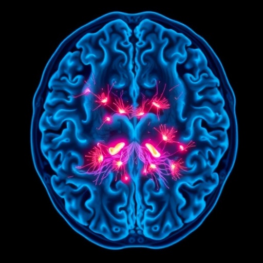
Scientists have captured a seizure moving through a living brain in three dimensions and in real time, offering one of the clearest views yet of how abnormal electrical activity spreads through neural tissue. Using a newly developed high-resolution light-sheet microscope, researchers at the University of Georgia recorded the progression of a seizure in a zebrafish larva from its apparent origin to its eventual disappearance. The advance could help neuroscientists understand how seizures begin, why they travel through particular brain regions and how they might be stopped before they develop into more serious neurological events.
A seizure is often described as a sudden burst of uncontrolled electrical activity in the brain, but that description conceals an extraordinarily complex process. Within seconds, networks of neurons can shift from coordinated signaling into a state of excessive, synchronized activity. The event may begin in a small region and then move across interconnected areas, or it may recruit several regions almost simultaneously. Because the activity develops so quickly and inside a three-dimensional organ, researchers have struggled to observe the entire event with sufficient spatial and temporal detail. Conventional imaging methods can show activity across a flat plane, but they may miss what is happening above or below that slice.
The University of Georgia team addressed this problem by combining light-sheet microscopy with adaptive optics, creating an imaging system capable of capturing rapid changes throughout the brain of a living zebrafish larva. In light-sheet microscopy, a thin plane of laser light illuminates only one narrow slice of the specimen at a time. Fluorescent signals produced by active neurons can then be recorded by a camera positioned perpendicular to the light sheet. By rapidly moving the illuminated plane through the sample and assembling the resulting images, the microscope generates a volumetric view of neural activity. The approach limits unnecessary illumination and reduces background noise, allowing researchers to image delicate living organisms at high speed.
The system’s adaptive optics component further improves the quality of the images. When light passes through biological tissue, it encounters structures with different optical properties. Those variations bend and scatter the light, distorting the signal before it reaches the microscope. The result is a blurred image, particularly when researchers attempt to look deep into tissue or across an extended volume. Adaptive optics corrects these distortions by measuring how the light has been altered and adjusting the optical system to compensate. The technology was originally developed for astronomy, where it is used to correct the blurring caused by Earth’s atmosphere and produce sharper images of distant stars and galaxies.
In the new experiments, zebrafish larvae served as a transparent and experimentally accessible model for studying seizure activity. Zebrafish are widely used in neuroscience because their early-stage bodies are small, their nervous systems share important biological features with those of other vertebrates and their developing tissues can often be observed with minimal obstruction. Their transparency is especially valuable for optical imaging, making it possible to monitor activity across the brain without the extensive surgical procedures required in many mammalian models. The larvae also provide a compact system in which researchers can follow the movement of electrical activity across multiple brain regions at once.
The recorded seizure appeared to begin toward the rear of the larval brain before advancing toward the front. As the activity moved forward, it reached the optic tectum, a midbrain structure involved in processing visual information. In zebrafish, the optic tectum plays a central role in interpreting what the animal sees, coordinating eye movements and generating behavioral responses to visual stimuli. The imaging sequence showed that the abnormal activity did not remain confined to a single location. Instead, it propagated through the brain in a spatially organized pattern before gradually weakening over several seconds. The resulting footage provides a rare start-to-finish view of a seizure spreading through a living vertebrate brain.
That sequence is significant because seizure propagation is not merely a visual phenomenon; it reflects the organization of the underlying neural circuits. Neurons communicate through electrical and chemical signals, and their connections determine how activity can travel from one population to another. During a seizure, normal regulatory mechanisms that prevent excessive excitation can become overwhelmed or disrupted. Mapping the route of the event in three dimensions may help researchers identify which circuits act as launch points, which regions amplify the activity and which areas limit its spread. Such information could eventually contribute to more precisely targeted treatments for epilepsy and other disorders involving abnormal brain excitability.
Peter Kner, a professor in the University of Georgia’s College of Engineering and the study’s corresponding author, emphasized the limitations of relying on two-dimensional images to understand a three-dimensional brain. A single optical plane may show a wave of activity moving across the field of view, but it cannot establish whether the wave continues outside that plane or whether apparently separate signals are connected at another depth. Volumetric imaging reduces that uncertainty by allowing researchers to track activity across the full thickness of the brain. The added dimension can reveal pathways, timing relationships and patterns of recruitment that might otherwise be mistaken for isolated events.
The microscope could also become useful beyond seizure research. High-speed, low-background light-sheet imaging is suited to observing many fast biological processes, including the development of neural circuits, the movement of immune cells and changes in blood flow. Adaptive optics may allow researchers to examine these events more sharply in thicker or more optically complex tissues. For neuroscience, the combination could help connect cellular activity with behavior by showing how neural networks operate while an organism responds to its surroundings. Although findings from zebrafish cannot be transferred directly to human patients, the model offers a practical way to test biological mechanisms and potential therapies before they are studied in more complex systems.
The study, published in Biomedical Optics Express, demonstrates how advances in optical engineering can change the questions researchers are able to ask about the brain. The work was co-authored by Bingxi Liu, Yang Liu, Carly Duffy and James Lauderdale, and was supported by the National Institutes of Health. By recording a seizure as a rapidly evolving three-dimensional event rather than as a signal on a flat screen, the researchers have brought scientists closer to understanding how pathological activity takes shape and travels through neural networks. The next challenge will be to use this detailed view to compare different seizure types, identify the mechanisms that terminate them and determine whether specific points along their route can be targeted to prevent their spread.
News Publication Date: 21 July 2026
Web References: Biomedical Optics Express article; Peter Kner, University of Georgia; James Lauderdale, University of Georgia
References: Biomedical Optics Express, DOI: 10.1364/BOE.596096
Keywords
Seizures, epilepsy research, zebrafish neuroscience, brain imaging, light-sheet microscopy, adaptive optics, volumetric imaging, seizure propagation, biomedical engineering, neural activity
Cite Scienmag News
Cassandra Pierce. (August 13, 2026). New Imaging Technology Captures Seizures as They Happen. Scienmag. https://scienmag.com/new-imaging-technology-captures-seizures-as-they-happen/
Cassandra Pierce. "New Imaging Technology Captures Seizures as They Happen." Scienmag, 13 August 2026, https://scienmag.com/new-imaging-technology-captures-seizures-as-they-happen/. Accessed 4 September 2026.
Cassandra Pierce. "New Imaging Technology Captures Seizures as They Happen." Scienmag. August 13, 2026. https://scienmag.com/new-imaging-technology-captures-seizures-as-they-happen/


