


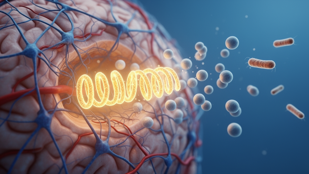
Pneumonia is one of the most feared complications after an ischemic stroke. Roughly one in three stroke patients develops an infection during their hospital stay, and pneumonia alone strikes between 8.5 and 14.3 percent of them, rising to 28 percent among those in intensive care. Clinicians have long observed that patients who catch pneumonia fare worse, but the biological reasons have remained frustratingly opaque. Now, a team led by Dirk M. Hermann at University Hospital Essen, working within the German Research Foundation’s Collaborative Research Center on neutrophil biology, has traced the damage to a surprising culprit: positively charged histone proteins released by overstimulated neutrophils. Their study, published in Acta Neuropathologica, shows that antibody-mediated neutralization of extracellular histones can rescue neurological recovery and prevent long-term brain atrophy in mice with stroke-associated pneumonia, even when antibiotics cannot.
The investigation began in the clinic. Within the prospective NOFF-S cohort, recruited through the Stroke Unit of the University Hospital Essen since October 2022, the researchers compared 26 patients who developed stroke-associated pneumonia with 326 who did not. Patients with pneumonia had more severe strokes at admission, measured by the National Institutes of Health Stroke Scale, and their 90-day outcome on the modified Rankin Scale was markedly worse. Pneumonia predicted poor outcome with a relative risk of 3.2 in both univariate and multivariable analyses, and the risk only modestly declined after adjustment for age, sex, and initial stroke severity. Flow cytometry of blood samples revealed something equally striking: patients who developed pneumonia showed a long-lasting deregulation of neutrophil subsets, with reduced proportions of CD15-low neutrophils persisting even three months after the stroke, hinting that the immune disturbance triggered by pneumonia was not a transient event.
To dissect mechanisms, the team turned to mice. They induced a transient 30-minute occlusion of the middle cerebral artery and, three days later, instilled Streptococcus pneumoniae directly into the trachea, replicating the typical timing of pneumonia onset in stroke patients. The results were dramatic. Pneumonia did not enlarge the initial infarct, but it significantly worsened neurological deficits, increased brain edema, and broke down the blood-brain barrier, as evidenced by increased IgG extravasation into the ischemic tissue. It also amplified the expression of the adhesion molecule ICAM-1 on cerebral microvessels, boosted infiltration of CD45-positive leukocytes, increased the density of platelet-rich microvascular thrombi marked by glycoprotein-Ib-alpha, and left behind extranuclear deposits of histone-H4 in the previously ischemic striatum, including in microglia and macrophages. A more severe 60-minute stroke model reproduced the same pattern, confirming that the findings were not an artifact of mild ischemia.
Perhaps the most sobering result concerned standard care. Amoxicillin, started three hours after infection and repeated every eight hours, reduced the bacterial load in the lungs and partially restored blood-brain barrier integrity and microvascular thrombosis. Yet it failed to improve neurological deficits, did not reduce the flood of neutrophils, monocytes, T cells, B cells, and NK cells invading the ischemic brain, and left extranuclear histone deposits untouched. This mirrors the disappointing record of prophylactic antibiotics in large randomized stroke trials such as STROKE-INF and PASS, which consistently failed to improve outcome despite reducing infection rates. Infection control, the study makes clear, is not the same as controlling the inflammatory fallout of infection.
Proteomic analysis of sorted blood neutrophils revealed why. Among 1,058 detected proteins, pneumonia shifted the neutrophil proteome toward a signature of degranulation, platelet activation, and NETosis, the explosive process by which neutrophils eject web-like structures of DNA studded with antimicrobial and tissue-damaging proteins. Twelve proteins were regulated in opposite directions by pneumonia and amoxicillin, including the chromatin-remodeling protein RUVBL1 and the histone H2B family member HIST2H2BB, interpreted as markers of ongoing NETosis and prior histone release. Notably, TOLLIP, a positive regulator of chronic inflammation known to exacerbate ischemic brain injury, rose with pneumonia and resisted amoxicillin, indicating a proinflammatory program that antibiotics alone cannot switch off.
With neutrophils implicated, the researchers tested a series of interventions. Depleting neutrophils with an anti-Ly6G antibody reversed the pneumonia-driven blood-brain barrier breakdown, brain edema, and microvascular thrombosis, establishing neutrophils as central mediators. But neutrophils are also essential for bacterial clearance, and post-stroke immunosuppression already compromises adaptive immunity, so depletion is not a clinically viable option. The team therefore targeted neutrophil products instead. Degrading existing neutrophil extracellular traps with DNase-I did nothing to help and actually increased hemorrhagic transformation of the infarcts, suggesting that NET DNA helps seal fragile ischemic vessels. Blocking NET formation with the gasdermin-D inhibitor LDC7559, or inhibiting the NET-bound enzyme myeloperoxidase with AZD4831, each reduced brain edema and microvascular thrombosis, but neither restored neurological function when given at the time of pneumonia.
Only one strategy delivered the complete package. A monoclonal antibody, BWA3, that neutralizes extracellular histone-H4 and H2A, administered just before and one day after pneumonia induction, reduced neurological deficits, shrank infarct volume, prevented blood-brain barrier breakdown, and curtailed microvascular thrombosis. Crucially, the benefits endured. Over 56 days of follow-up, histone-neutralized mice performed better on neurological scoring, tight rope walking, open field activity, and novel object recognition, and they developed significantly less brain atrophy. Histone blockade also increased the density of microglia and macrophages in the ischemic striatum while reducing their activation, without affecting microglial cell death, and it lowered circulating extracellular histone-H4 without altering NET levels, indicating that the antibody disarms histones rather than preventing their release.
The biology behind this protection fits a growing picture of extracellular histones as potent damage-associated molecular patterns. Once outside the cell, histones injure tissue by activating toll-like receptors and the complement cascade, triggering cytokine and chemokine release through MyD88, nuclear factor-kappa-B, and the NLRP3 inflammasome. In atherosclerosis models, externalized histone-H4 kills vascular smooth muscle cells, recruits more neutrophils, and perpetuates a cycle of NET release that destabilizes plaques, a cycle that histone neutralization can break. Extracellular histones have previously been identified as major mediators of death in sepsis, and administering histones to stroke mice worsens neurological deficits and infarct volume. What is new here is the demonstration that histones are the decisive link between a lung infection and progressive brain injury, and that intercepting them days after the stroke, at the moment pneumonia strikes, can still change the trajectory of recovery.
The clinical implications are tantalizing. Small-molecule inhibitors, inhibitory peptides, and neutralizing antibodies directed against histone-H4 are already in development for other inflammatory conditions, and the new data argue that stroke patients developing pneumonia could be a prime population for such therapies. The study also carries a caution for ongoing trials of intravenous DNase-I in ischemic stroke, three of which are currently registered, since delayed NET degradation increased brain hemorrhages in this model and should be monitored carefully. For now, the work redefines stroke-associated pneumonia not simply as an infection to be cured but as a neutrophil-driven storm whose toxic cargo, the histones, can be intercepted. If the mouse findings translate, the humble proteins that package DNA in every cell nucleus may become one of the most actionable drug targets in post-stroke care.
Subject of Research: Neutrophil-driven inflammatory brain injury and histone neutralization therapy in ischemic stroke complicated by bacterial pneumonia
Article Title: Histone neutralization protects the ischemic brain against the consequences of stroke-associated pneumonia
Article References: Yin, D., Li, A., Mohamud Yusuf, A., Shevchuk, O., Gronewold, J., Thiebes, S., Tertel, T., Wang, C., Hagemann, N., Zhang, Y., Graser, C., Tas, H., Fleischer, M., Kaltwasser, B., Frank, B., Tuz, A. A., Singh, V., Siemes, D., Pylaeva, E., … Hermann, D. M. (2026). Histone neutralization protects the ischemic brain against the consequences of stroke-associated pneumonia. Acta Neuropathologica, 152(1), Article 50. https://doi.org/10.1007/s00401-026-03097-0
Image Credits: AI Generated
DOI: 10.1007/s00401-026-03097-0
Keywords: ischemic stroke, stroke-associated pneumonia, neutrophils, neutrophil extracellular traps, extracellular histones, histone neutralization, blood-brain barrier, microvascular thrombosis, Streptococcus pneumoniae, myeloperoxidase, brain atrophy, neurological recovery
Cite Scienmag News
Cassandra Pierce. (October 9, 2026). Neutralizing Histones Shields the Stroke-Injured Brain from Pneumonia Damage. Scienmag. https://scienmag.com/neutralizing-histones-shields-the-stroke-injured-brain-from-pneumonia-damage/
Cassandra Pierce. "Neutralizing Histones Shields the Stroke-Injured Brain from Pneumonia Damage." Scienmag, 9 October 2026, https://scienmag.com/neutralizing-histones-shields-the-stroke-injured-brain-from-pneumonia-damage/. Accessed 9 October 2026.
Cassandra Pierce. "Neutralizing Histones Shields the Stroke-Injured Brain from Pneumonia Damage." Scienmag. October 9, 2026. https://scienmag.com/neutralizing-histones-shields-the-stroke-injured-brain-from-pneumonia-damage/


