


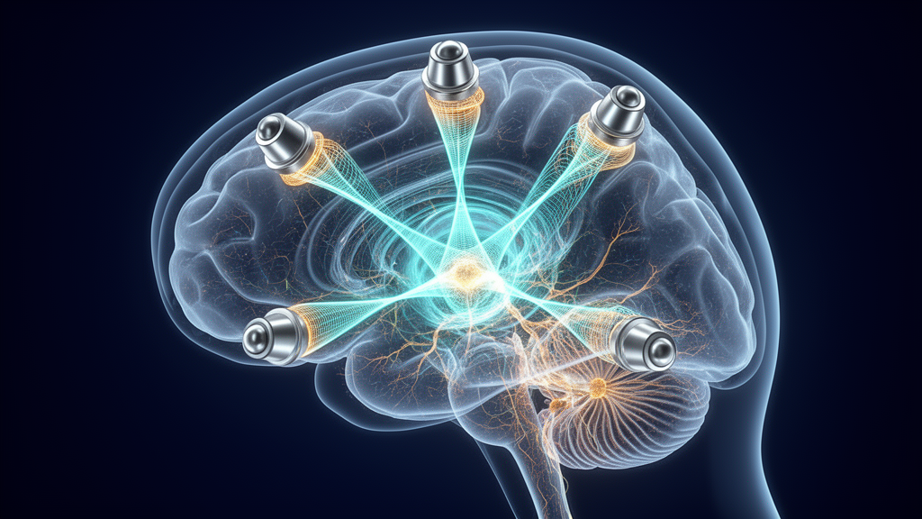
Noninvasive electrical brain stimulation offers a compelling path for exploring neural circuits and designing interventions for neurological conditions, yet standard transcranial modalities have struggled to target deep anatomical structures without unintentionally altering superficial cerebral cortex. Conventional approaches such as transcranial electrical stimulation and transcranial magnetic stimulation deliver their strongest fields directly beneath the scalp, causing physical field strength to decay rapidly with anatomical depth. To bypass this limitation, temporal interference stimulation was previously introduced as a noninvasive alternative, delivering pairs of high-frequency kilohertz electric fields with small frequency offsets through scalp electrodes. These high-frequency carrier currents pass relatively harmlessly through intervening cortical tissue before intersecting at a subcortical target, where temporal interference generates a low-frequency amplitude-modulated envelope capable of driving neural responses. However, conventional temporal interference has remained constrained by a rigid physical trade-off: researchers could not adjust the spatial focality of the stimulated area independently from the intensity of the stimulation envelope, because turning up the stimulation current inherently expands the surrounding volume of activated tissue.
To overcome this long-standing engineering hurdle, an international research team led by Boris Botzanowski, Brian D. Corneil, and Adam Williamson developed multipolar temporal interference, an expanded stimulation architecture designed to decouple the physical volume of activation from the delivered stimulation amplitude. Rather than relying on a single pair of interfering kilohertz carrier waves to generate a solitary amplitude-modulated envelope, the multipolar configuration superimposes multiple independent carrier pairs, each operating at distinct baseline frequencies but tuned to share an identical envelope difference frequency. By spacing the average frequency of each carrier pair at least one kilohertz apart, the protocol prevents unintended cross-channel beat frequencies from producing spurious low-frequency envelopes outside the selected target structure. When the stimulating currents across these multiple electrode sets are scaled down, the overlapping electrical fields sum together at the anatomical convergence point to match the envelope intensity of standard temporal interference while sharply contracting the surrounding field distribution.
The physical mechanisms governing this spatial sharpening were first validated through computational finite element method simulations performed on the Sim4Life platform using a detailed anatomical model of a rhesus macaque head. Solving the Laplace equation across more than three hundred and fifty segmented tissues under quasi-electrostatic ohmic approximations, the researchers mapped the spatial field distributions generated by conventional two-frequency temporal interference against the newly developed multipolar temporal interference paradigm. Setting an activation threshold at six volts per meter, the computational simulations revealed that multipolar temporal interference contracted the volume of activated tissue into a significantly tighter envelope centered on the target structure. Cumulative histogram evaluations demonstrated that with multipolar temporal interference, less than one percent of non-target brain volume received an electric field higher than the targeted region, whereas standard two-channel temporal interference exposed more than twenty percent of the surrounding brain tissue to field intensities exceeding those delivered to the target.
To evaluate these theoretical predictions in living tissue, the investigators first tested the method in an anesthetized male rhesus macaque instrumented with sixteen transcutaneous stimulation electrodes and stereoelectroencephalography depth probes. The recording setup comprised nine discrete burr-hole craniotomies guiding multielectrode depth shafts across temporal lobe structures, enabling spatial sampling of intracranial potentials at fifteen contacts per shaft. Multipolar temporal interference was delivered using eight current stimulators driving four distinct carrier pairs centered at difference frequencies generating fifty-hertz envelopes, with individual current intensities ranging from 1.1 to 2.6 milliamperes to establish a uniform 750-microvolt peak-to-peak envelope amplitude at the deep reference contact. Calculating the modulation index across intracranial recording sites demonstrated that while conventional two-carrier stimulation generated broad, diffuse subcortical modulation, the eight-carrier multipolar approach produced a markedly more localized envelope maximum centered precisely on the monitored target electrode.
The researchers subsequently tested whether the focality improvements of multipolar temporal interference scale downward into much smaller mammalian anatomical dimensions by conducting parallel experiments in anesthetized mice. Using twelve-week-old OF1 mice, the team surgically placed eight pairs of minimally invasive stimulation screws in a radial array surrounding a thirty-two-channel linear silicon depth probe inserted directly through the hippocampus. Standard temporal interference delivered two thousand microamperes per electrode pair using two carrier frequencies, whereas multipolar temporal interference delivered two hundred and fifty microamperes per pair across eight carrier frequencies, both adjusted to produce an artifact amplitude of approximately thirty-two hundred microvolts at target contact sixteen. Electrophysiological recordings revealed that conventional temporal interference produced little spatial variation in modulation index across the entire recording track, whereas multipolar temporal interference generated a prominent, sharp peak centered at the hippocampal target that diminished steeply across adjacent dorsal and ventral contacts.
Moving beyond anesthetized preparations, the team assessed whether multipolar temporal interference could selectively modulate electrophysiological firing patterns within the midbrain of an awake, behaving male rhesus macaque performing structured oculomotor tasks. The investigators targeted the intermediate layers of the superior colliculus, a midbrain structure that plays a well-characterized functional role in commanding gaze shifts and saccadic eye movements. Using a sixteen-contact laminar S-probe lowered through an acrylic chamber, the researchers isolated neurons that exhibited functional discharges during visually guided saccades. Transcutaneous stimulation was applied through sixteen scalp electrodes arranged around the cranial implant, delivering a burst of four overlapping high-frequency carrier pairs spanning from approximately two to eight kilohertz, offset to create an aggregate one-hundred-hertz modulation envelope measuring four hundred microvolts at the selected intermediate collicular contact.
In the first behavioral paradigm, the monkey maintained prolonged visual fixation on a central target for 1900 to 2300 milliseconds, during which a two-hundred-millisecond train of multipolar temporal interference or sham stimulation was delivered during the middle of the fixation window. Because the high-amplitude electrical carriers saturated recording amplifiers during active delivery, neural spike counts were quantified immediately following stimulation offset, spanning an evaluation window between 250 and 350 milliseconds after stimulation onset. Across fifteen single units recorded simultaneously during this fixation task, nine neurons displayed statistically significant changes in post-stimulation firing rates relative to non-stimulated control trials, six neurons showed significant firing differences compared to sham stimulation trials where carrier frequencies lacked offset, and only two neurons showed significant changes between sham and baseline conditions under Bonferroni-corrected Wilcoxon rank-sum testing.
The investigators next evaluated whether multipolar temporal interference could modulate functionally recruited neural activity during active movement preparation in a visually guided saccade task. In this second paradigm, the fixation point vanished for a one-hundred-millisecond gap before a peripheral target appeared, with two-hundred-millisecond stimulation trains timed to terminate approximately thirty milliseconds after target appearance, just preceding the arrival of endogenous visual signals in the superior colliculus. Across fourteen functionally identified saccade-related neurons, four units demonstrated statistically significant firing rate elevations during a post-target window spanning forty to one hundred milliseconds compared to both no-stimulation and sham controls. At the population level, a paired t-test confirmed that average firing rates across all fourteen recorded neurons rose to 111.7 hertz following multipolar stimulation, significantly exceeding the 96.5 hertz observed during unmodulated baseline trials and the 100.1 hertz measured during carrier-matched sham trials.
Behavioral monitoring during both tasks showed that transcutaneous multipolar temporal interference did not disrupt the monkey’s task execution, alter gaze accuracy, or evoke visible motor twitches, blinking, or signs of distress. In the prolonged fixation task, delivering hundred-hertz multipolar stimulation did not trigger premature saccades away from the fixation point, confirming that the stimulation remained below the threshold required to force overt motor outputs. However, the multipolar stimulation trains did elicit measurable pupil dilation during the fixation window, a physiological reaction consistent with subthreshold intermediate superior colliculus activation documented in prior intracortical microstimulation studies. Sham stimulation also produced pupil dilation, although to a lesser magnitude, which the researchers noted highlights the necessity of refining sham protocols to control for cutaneous sensory percepts that can arise when switching unramped kilohertz carrier currents across scalp electrodes.
The study also provides practical technical insights into selecting frequency separations when designing multichannel noninvasive neuromodulation arrays. Previous attempts to improve temporal interference focality by adding secondary electrode channels retained identical carrier frequencies across pairs, an arrangement that enlarges the aggregate envelope volume rather than contracting it. By enforcing an inter-pair frequency separation of at least one kilohertz, the authors demonstrated that high-frequency cross-channel mixing products fall well above physiological cellular integration bandwidths, preventing unwanted modulation outside the focal intersection. In human cortical exposure work referenced by the authors, currents delivered at two thousand hertz exhibited dramatically higher activation thresholds than one-thousand-hertz exposures, supporting the researchers’ decision to separate active carrier groups by up to two thousand hertz in their awake macaque protocol to suppress peripheral side effects.
Notwithstanding these electrophysiological demonstrations, the authors acknowledged several methodological limitations and outlined critical directions for subsequent research before clinical translation can occur. The awake neurophysiological data were obtained from a single nonhuman primate performing specific oculomotor tasks across two recording days, underscoring the need to reproduce these deep-brain modulation effects across larger cohorts and multiple behavioral contexts. Furthermore, because high-frequency carrier currents overwhelmed recording amplifiers during active delivery, immediate intra-stimulation cellular dynamics could not be directly observed, restricting analyses to the period following stimulation offset. Future human applications will also require systematic psychophysical assessments to characterize scalp sensations and implement ramped sham controls, alongside high-resolution individualized finite element models to optimize electrode placement across varied cranial geometries.
Subject of Research: Focal noninvasive deep brain stimulation using multipolar temporal interference
Article Title: Focal control of non-invasive deep brain stimulation using multipolar temporal interference
Article References: Botzanowski, B., Acerbo, E., Lehmann, S., Kearsley, S. L., Steiner, M., Neufeld, E., Missey, F., Muller, L., Jirsa, V., Corneil, B. D., & Williamson, A. (2025). Focal control of non-invasive deep brain stimulation using multipolar temporal interference. Bioelectronic Medicine, 11(1), Article 7. https://doi.org/10.1186/s42234-025-00169-6
Image Credits: AI Generated
DOI: 10.1186/s42234-025-00169-6
Keywords: Temporal Interference, Multipolar Temporal Interference, Deep Brain Stimulation, Noninvasive Neuromodulation, Superior Colliculus, Rhesus Macaque, Electrophysiology, Hippocampus, Bioelectronic Medicine, Transcranial Stimulation, Finite Element Modeling, Neural Spiking, Saccadic Eye Movements, Stereoelectroencephalography, Pupil Dilation, Modulation Index
Cite Scienmag News
Cassandra Pierce. (November 11, 2026). Multipolar Temporal Interference Sharpens Noninvasive Deep Brain Stimulation in Animal Models. Scienmag. https://scienmag.com/multipolar-temporal-interference-sharpens-noninvasive-deep-brain-stimulation-in-animal-models/
Cassandra Pierce. "Multipolar Temporal Interference Sharpens Noninvasive Deep Brain Stimulation in Animal Models." Scienmag, 11 November 2026, https://scienmag.com/multipolar-temporal-interference-sharpens-noninvasive-deep-brain-stimulation-in-animal-models/. Accessed 11 November 2026.
Cassandra Pierce. "Multipolar Temporal Interference Sharpens Noninvasive Deep Brain Stimulation in Animal Models." Scienmag. November 11, 2026. https://scienmag.com/multipolar-temporal-interference-sharpens-noninvasive-deep-brain-stimulation-in-animal-models/




