


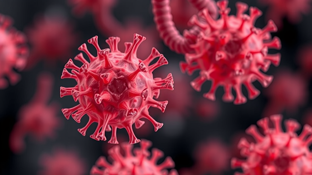
The community of bacteria living deep within the lungs, long regarded as sparse and relatively inconsequential compared with the dense microbial ecosystems of the gut, is emerging as a decisive factor in how severe an influenza infection becomes. A new study published in the journal Microbiome reports that when the lung microbiota falls into a state of dysbiosis, a disruption of its normal composition, the imbalance can actively worsen infection with influenza A virus (IAV). Working with mouse models that reproduce different levels of disease severity, researchers traced the damaging effect to a single opportunistic pathogen, Bacillus cereus, and to a specific metabolic molecule it releases that quietly disarms one of the lung’s most important antiviral defenses.
The research team, led by Hai Chang Yin, Xin Yu Zhang and Song Liu of Qiqihar University together with colleagues at the Harbin Veterinary Research Institute and the Heilongjiang Academy of Agricultural Sciences, set out to map the relationships between pulmonary microbes, their metabolites and the host immune response during IAV infection. Prior work had established that microbes in the respiratory tract help shape host immunity and reduce the risk of respiratory infections, but the mechanisms by which an unbalanced lung community might aggravate viral disease remained poorly defined. By establishing models of mild and severe influenza disease in mice and analyzing how the pulmonary microbiota changed alongside the host response, the investigators found that the microbial shifts were characterized by the enrichment of opportunistic pathogenic groups, and that the degree of that enrichment tracked with disease severity.
Multi-omics analysis, which integrates sequencing data on microbial communities with measurements of metabolites and host gene expression, pointed the investigators toward xanthosine, a purine metabolite. According to the study, colonization of the lungs by Bacillus cereus aggravates IAV infection through this molecule. Xanthosine, the researchers determined, downregulates the expression of nt5e, a gene encoding ecto-5′-nucleotidase, also known as CD73, an enzyme stationed on the surface of alveolar macrophages. The consequence of suppressing nt5e is a reduction in the viral phagocytic ability of these macrophages, the resident immune cells that patrol the air sacs of the lungs and engulf virus particles and infected cellular debris. In effect, the bacterial metabolite blinds and slows the very cells responsible for clearing the virus, allowing influenza to replicate more freely and inflict greater damage on lung tissue.
The finding reframes the interaction between bacteria and viruses in the respiratory tract. Rather than merely serving as passive bystanders or secondary invaders that exploit virus-damaged tissue, certain lung bacteria can actively promote viral pathogenesis through chemical signaling. The xanthosine-nt5e axis identified in this study offers a concrete molecular link between microbial dysbiosis and impaired innate immunity. Because alveolar macrophages represent a first line of defense against inhaled pathogens, their impairment early in infection can shape the entire trajectory of disease, determining whether an influenza infection remains mild or spirals into severe pneumonia. The severity-dependent patterns observed across the mouse models suggest that this mechanism operates along a gradient: the more pronounced the dysbiosis and the greater the enrichment of opportunistic pathogens such as B. cereus, the weaker the macrophage response and the worse the outcome.
Perhaps the most consequential part of the study is its demonstration that the damage is reversible. Using bacteriophages, viruses that infect and kill specific bacteria, the researchers targeted and removed B. cereus from the lungs of infected mice. This phage-mediated clearance alleviated IAV infection and reduced the resulting pulmonary lesions. In other words, precisely editing the lung microbiota, rather than broadly suppressing bacteria with antibiotics, was enough to restore a meaningful degree of antiviral protection. The approach sidesteps a well-known problem with conventional antibiotics, which can indiscriminately disturb beneficial communities and select for resistance, and instead offers a scalpel-like intervention aimed at a single harmful player.
The technical architecture of the study deserves attention. The team did not rely on a single measurement platform; instead, the multi-omics workflow connected three layers of biology: which bacterial groups expanded during infection, which metabolites those groups produced, and which host genes and immune functions changed in response. Animal experiments then validated the causal chain implied by the correlational data. By showing that B. cereus colonization, xanthosine signaling, nt5e suppression and macrophage dysfunction form a coherent mechanistic pathway, and that interrupting the pathway at its origin through phage therapy rescues the host, the study satisfies a standard of evidence that many microbiome associations lack. The work was approved by Qiqihar University and conducted under animal ethics guidelines and approved protocols.
The implications extend beyond influenza. Influenza A virus remains one of the most consequential respiratory pathogens globally, causing seasonal epidemics and occasional pandemics, and severe outcomes are often driven by immunopathology in the lower respiratory tract. If a comparable dysbiosis-driven mechanism operates in humans, the composition of a patient’s lung microbiota could serve as a biomarker for predicting who is most likely to develop severe disease, and microbiota-targeted interventions could become part of the clinical toolkit alongside antivirals and vaccines. The authors conclude that their findings highlight a novel approach for preventing and controlling the progression of IAV infection by targeting the lung microbiota itself, a strategy that complements rather than replaces existing countermeasures.
Several questions will need to be answered before such a strategy reaches the clinic. The study was performed in specific pathogen-free mice, and the human lung microbiome differs in composition and density from that of laboratory animals. Whether B. cereus or related opportunists occupy a comparable niche in human airways during influenza, and whether xanthosine signaling suppresses macrophage function through the same nt5e-dependent route in patients, remain open issues. Distinguishing cause from consequence in severely ill patients will also be critical, since viral tissue damage can itself reshape the microbial environment. Nonetheless, the concept that a bacterial metabolite can act as an immunological saboteur during viral infection is a testable and therapeutically attractive hypothesis.
The phage-based rescue demonstrated in the mice suggests one immediate translational path. Bacteriophage therapy has attracted renewed interest as antimicrobial resistance spreads, and this study adds a virology-adjacent application: using phages not to treat bacterial disease directly, but to correct a microbial imbalance that is worsening a viral one. As sequencing technologies make it faster and cheaper to profile respiratory microbiomes, clinicians could eventually identify high-risk dysbiotic states at admission and deploy targeted phage cocktails to remove specific aggravating species before viral loads climb. Such an approach would represent a genuinely new layer of precision medicine for respiratory infections, one that treats the microbial ecosystem as an active participant in disease rather than scenery around it.
For now, the study stands as a clear demonstration that the boundaries between bacterial and viral disease are more porous than traditional thinking assumed. A metabolite released by an opportunistic lung bacterium can reach across the species divide, dampen a key antiviral enzyme on immune cells, and tip the balance of an influenza infection toward severity. That the process can be reversed by removing a single bacterial species with phages gives the finding practical weight. It also adds to a growing body of evidence that maintaining a healthy respiratory microbiota, or actively repairing a damaged one, may be as important to defending the lung against influenza as any single antiviral drug.
Subject of Research: How lung microbiota dysbiosis, specifically Bacillus cereus and its metabolite xanthosine, exacerbates influenza A virus infection by impairing alveolar macrophage function
Article Title: Lung microbiota dysbiosis exacerbates influenza virus infection
Article References: Yin, H. C., Zhang, X. Y., Liu, S., Jiang, X., Yu, T. F., Zhang, H., Liu, D., & Xia, C. Y. (2026). Lung microbiota dysbiosis exacerbates influenza virus infection. Microbiome. https://doi.org/10.1186/s40168-026-02450-5
Image Credits: AI Generated
DOI: 10.1186/s40168-026-02450-5
Keywords: lung microbiota, influenza A virus, dysbiosis, Bacillus cereus, xanthosine, nt5e, alveolar macrophages, bacteriophages, microbiome, respiratory infection, innate immunity, phage therapy
Cite Scienmag News
Kristina Jarvis. (September 21, 2026). Lung Bacteria Found to Worsen Influenza Through a Metabolic Trap in Immune Cells. Scienmag. https://scienmag.com/lung-bacteria-found-to-worsen-influenza-through-a-metabolic-trap-in-immune-cells/
Kristina Jarvis. "Lung Bacteria Found to Worsen Influenza Through a Metabolic Trap in Immune Cells." Scienmag, 21 September 2026, https://scienmag.com/lung-bacteria-found-to-worsen-influenza-through-a-metabolic-trap-in-immune-cells/. Accessed 5 October 2026.
Kristina Jarvis. "Lung Bacteria Found to Worsen Influenza Through a Metabolic Trap in Immune Cells." Scienmag. September 21, 2026. https://scienmag.com/lung-bacteria-found-to-worsen-influenza-through-a-metabolic-trap-in-immune-cells/


