


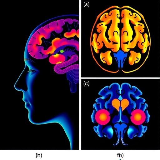
Depression may not have a single neural signature shared equally by men and women. A large-scale brain-imaging study of 853 young adults has found that depression was associated with reduced activity in different parts of the prefrontal cortex depending on sex. In men with depression, the most prominent alteration appeared in the right frontal eye field, a region involved in directing attention and controlling eye movements. In women with depression, reduced activity was concentrated in the left dorsolateral prefrontal cortex, an area central to working memory, cognitive control, decision-making and emotional regulation. The findings challenge the increasingly outdated idea that depression can be represented by one generalized pattern of “underactive” frontal brain activity.
The study, published in Annals of General Psychiatry, examined 431 people diagnosed with depression and 422 healthy controls. Participants were between 18 and 45 years old, right-handed, and recruited through the psychiatry department of an affiliated university hospital in China. Three senior psychiatrists confirmed depression diagnoses using structured clinical interviews based on the Diagnostic and Statistical Manual of Mental Disorders, Fifth Edition. The researchers also used the Hospital Anxiety and Depression Scale to quantify depressive symptoms. The depression group scored substantially higher than the healthy group, while the two groups did not differ significantly in age. Participants with other psychiatric disorders, major medical illnesses, substance-use problems, brain injuries or severe cognitive limitations were excluded.
To observe the brain while it was working, the researchers used functional near-infrared spectroscopy, or fNIRS. Unlike magnetic resonance imaging, fNIRS does not require a large scanner. Instead, a cap-like array places light emitters and detectors on the scalp. Near-infrared light at wavelengths of 760 and 850 nanometres penetrates the skull and is partly absorbed by blood oxygen-carrying molecules. When a brain region becomes more engaged, local blood flow generally increases, changing the relative concentrations of oxygenated and deoxygenated haemoglobin. The instrument recorded these changes across 53 channels covering the frontal poles, bilateral dorsolateral prefrontal regions and frontopolar cortex. Because oxygenated haemoglobin typically provides a stronger task-related signal, the researchers focused their main analysis on changes in oxyhaemoglobin.
The participants completed a verbal fluency task, a deceptively simple test that places heavy demands on executive control. After a 30-second counting period, each person heard a Chinese character and had 60 seconds to produce as many accurate two-character words as possible. They then completed a 60-second recovery period while counting aloud. Generating words under time pressure requires the brain to retrieve information from memory, suppress inappropriate responses, shift between possible answers and maintain attention on the rules of the task. These processes depend heavily on frontoparietal networks, particularly the dorsolateral prefrontal cortex. The task therefore offered the researchers a controlled way to examine how depression and sex interacted during cognitive effort rather than simply measuring brain activity at rest.
The researchers processed the optical recordings to remove physiological noise and movement-related distortions. Raw light intensities were converted into optical-density changes, and transient motion spikes were corrected with a wavelet-based method. A 0.01-to-0.10-hertz bandpass filter reduced interference from breathing, heartbeat and slow signal drift. Low-quality channels were removed when their coefficient of variation exceeded 15 percent during the task. The cleaned signals were then converted into haemoglobin concentration changes using the modified Beer–Lambert law, a mathematical model that estimates how light absorption relates to chromophore concentration in tissue. Task-related activation was calculated by comparing oxyhaemoglobin levels during the verbal fluency block with the preceding baseline.
The strongest sex-dependent pattern emerged in two channels corresponding to the left dorsolateral prefrontal cortex. Among participants with depression, women showed significantly lower oxyhaemoglobin activation than men in both channels. Within the female comparison, women with depression also showed lower activation in one of these channels than healthy women. The statistical interaction between sex and diagnostic group was marginally significant in the two left dorsolateral prefrontal channels, with probability values of 0.098 and 0.089, while the direct comparisons between depressed men and women were significant. These results point toward a particularly pronounced alteration in the left dorsolateral prefrontal cortex among women with depression, although the marginal interaction values mean the finding requires replication and should not be treated as a definitive biological boundary.
A different signal appeared in men. In the channel corresponding to the right frontal eye field, the interaction between sex and diagnostic group was statistically significant, with a probability value of 0.019. Men with depression showed lower activation in this region than healthy men, although the within-male comparison itself was described as marginally significant. The frontal eye field is best known for initiating voluntary and reflexive eye movements, including rapid saccades, but its role is broader than motor control. It forms part of the machinery that allocates spatial attention, selects relevant visual information and links perception to action. Reduced recruitment of this region could therefore reflect difficulties in shifting attention, searching efficiently or suppressing competing responses during cognitive activity. The result does not show that depression in men is caused by a malfunctioning eye-movement centre; rather, it identifies a region whose task-related blood-oxygen response differed between groups.
The left dorsolateral prefrontal cortex has a different functional profile. It maintains and manipulates information in working memory, helps form intentions, organizes goal-directed behaviour and contributes to the regulation of emotional information. It also communicates with sensory, premotor, parietal and limbic regions, making it a hub where thought, action and feeling can interact. Lower activation during verbal fluency could be consistent with the cognitive symptoms frequently reported in depression, such as slowed thinking, impaired concentration, difficulty making decisions and problems sustaining goal-directed behaviour. The researchers propose that the left dorsolateral prefrontal cortex may be a key location where the neural architecture of depression diverges between men and women. That interpretation is compatible with earlier neuroimaging studies linking depression to prefrontal dysfunction, but it remains an association rather than proof that the altered activity causes symptoms.
The results could eventually influence how researchers design treatments and biomarkers for depression, but they do not justify using a brain scan to diagnose an individual patient today. fNIRS is relatively inexpensive, portable and tolerant of natural movement, making it attractive for psychiatric research and potentially for repeated measurements during treatment. Yet the technique measures haemodynamic changes near the cortical surface, not electrical activity directly, and signals can be contaminated by blood flow in the scalp. The study did not use short-separation channels, which are designed to measure and regress out this superficial noise. It was also cross-sectional, so the researchers cannot determine whether the observed patterns precede depression, result from recurrent illness or change when symptoms improve. Most participants were young adults, and the task’s word-production performance was not analysed alongside the brain data. Future longitudinal studies spanning adolescence to older age, with detailed anxiety assessments, behavioural performance measures and improved physiological controls, will be needed to test whether sex-specific brain patterns can predict symptoms, treatment response or recovery.
Cite Scienmag News
Glenn Wilkins. (August 27, 2026). Large fNIRS study finds gender-specific prefrontal activation during verbal fluency in depression. Scienmag. https://scienmag.com/large-fnirs-study-finds-gender-specific-prefrontal-activation-during-verbal-fluency-in-depression/
Glenn Wilkins. "Large fNIRS study finds gender-specific prefrontal activation during verbal fluency in depression." Scienmag, 27 August 2026, https://scienmag.com/large-fnirs-study-finds-gender-specific-prefrontal-activation-during-verbal-fluency-in-depression/. Accessed 4 September 2026.
Glenn Wilkins. "Large fNIRS study finds gender-specific prefrontal activation during verbal fluency in depression." Scienmag. August 27, 2026. https://scienmag.com/large-fnirs-study-finds-gender-specific-prefrontal-activation-during-verbal-fluency-in-depression/


