


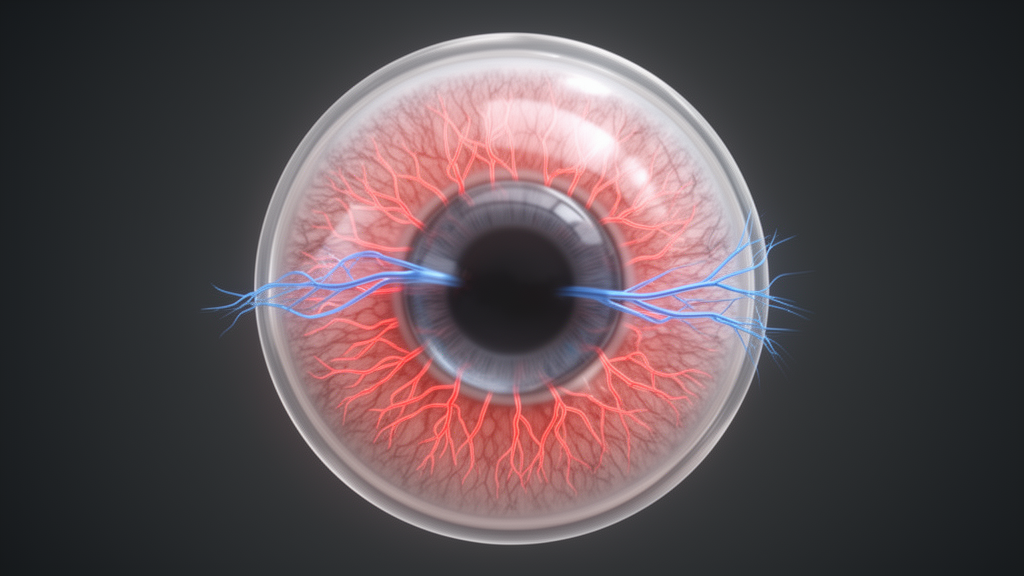
A 1–2-microliter injection of adeno-associated virus into the corneal stroma produced consistent labeling of neurons in the mouse trigeminal ganglion that project to the cornea, according to an experimental study comparing multiple viral serotypes, promoters and fluorescent reporters. The virus also enabled reporter expression in peripheral corneal nerves and in central brainstem projections. The findings establish a method for tracing and genetically accessing mouse corneal sensory neurons, not a clinical treatment or evidence of therapeutic benefit in people.
Corneal sensory fibers contribute to ocular-surface homeostasis and protection by detecting mechanical, thermal, chemical and osmotic stimuli and participating in responses such as blinking and tear secretion. Most corneal afferents are nociceptive. Altered corneal sensory signaling has also been implicated in neuropathic corneal pain, dry eye disease and neurotrophic keratopathy.
Conventional retrograde tracers can map the neurons supplying the cornea, but they cannot make those neurons express fluorescent sensors, optogenetic actuators, chemogenetic receptors, recombinases or other genetically encoded tools. The study examined whether intrastromal AAV delivery could provide that genetic access. Researchers working across independent laboratories injected viral vectors into the corneal stroma of mice ranging from five weeks to six months old.
They then examined cleared whole trigeminal ganglia, ganglion sections, corneal whole mounts and thick brainstem sections. Most assessments were conducted between two and 12 weeks after injection, while tissue collection extended as late as 22 weeks. The vector panel included AAV1, AAV2-7m8, AAV2-retro, AAV6, AAV8, AAV9 and AAV-PHP.S carrying eGFP, mCherry or tdTomato.
Cre-expressing AAV2-retro constructs were also tested in Ai14 tdTomato reporter mice to determine whether corneal delivery could activate a transgenic recombination-based labeling system. Across the tested constructs, neuronal labeling was concentrated in the dorsomedial part of the ophthalmic division of the trigeminal ganglion, corresponding to the expected location of cornea-projecting cell bodies. Some vector and reporter combinations labeled sparsely, while others produced many more labeled neurons.
AAV2-retro-CAG-tdTomato was among the strongest performers, and AAV6-CAG-eGFP also produced high labeling in sectioned tissue. Promoter and reporter selection influenced detection: in comparisons using AAV6, the CAG promoter generally supported more detectable neurons than CMV, while eGFP was more consistently detected than mCherry. In a separate serotype comparison, AAV-PHP.S labeled fewer neurons than AAV9 or AAV2-retro, whose labeling was comparable.
These findings show that vector choice cannot be treated as interchangeable across experimental settings. Fluorescent detection methods also changed the apparent extent of transduction. Native tdTomato fluorescence was readily detected in cleared ganglia, whereas antibody amplification strengthened eGFP signals, particularly in axonal fibers.
In one paired analysis, researchers identified an average of 74.3 eGFP-positive neurons using native fluorescence and 118.5 after anti-GFP immunolabeling, a significant difference. Among all detected neurons, 56% were visible with both methods, 40% only after antibody amplification and 4% only with native fluorescence. The comparison therefore indicates that direct viral fluorescence can undercount labeled neurons, particularly when eGFP is the reporter.
Sectioned and cleared whole-ganglion preparations gave similar overall results, although cleared preparations tended to contain more detectable cells. The researchers benchmarked viral labeling against conventional retrograde tracing from the cornea. When wheat germ agglutinin was injected into the cornea four weeks after AAV2-retro-CAG-tdTomato, AAV-labeled and tracer-labeled cell bodies occupied the same dorsomedial region of the ophthalmic division.
AAV labeling identified approximately 20% fewer neurons than wheat germ agglutinin, but the populations overlapped substantially: just over half of the combined labeled population expressed both markers, while nearly one-third was detectable only with the tracer. A separate Fast Blue comparison likewise found that most AAV-labeled neurons were also tracer-positive. The intentionally diluted vector in that experiment labeled roughly half as many neurons as Fast Blue, which the investigators attributed as the likely explanation for the reduced count.
The results support substantial correspondence between viral and conventional labeling rather than complete equivalence. Cellular markers and anatomical measurements were also consistent with corneal sensory neurons. About one-third of tdTomato-labeled neurons across three serotype and promoter combinations expressed calcitonin gene-related peptide, which is present in a subset of corneal afferents.
Soma-size distributions were compatible with a mixture of C-fiber and A-delta neurons. In mice whose NaV1.8-expressing nociceptors had been ablated, no AAV6-CAG-eGFP-labeled neurons were detected. Together, these observations support the interpretation that the vectors preferentially accessed nociceptive corneal sensory neurons, while leaving the broader immune profile unresolved.
Reporter expression extended from trigeminal cell bodies into peripheral and central portions of the sensory pathway. In corneal whole mounts, viral reporters appeared in stromal fibers, the sub-basal nerve plexus and intraepithelial terminals. Reporter-positive fibers in the sub-basal plexus were significantly associated with the number of labeled ganglion neurons, with an R-squared value of 0.72, F(1,4) equal to 10.4 and P equal to 0.03.
No significant relationship was detected for stromal or intraepithelial terminals. AAV2-retro produced greater tdTomato-labeled corneal nerve density than AAV9 or AAV-PHP.S. The investigators also developed a custom ImageJ plugin to separate stromal, sub-basal and intraepithelial nerve compartments during quantitative analysis, and used β-III-tubulin labeling to compare reporter expression with total corneal innervation. Central tracing showed that labeled axons entered the brainstem through the ventromedial trigeminal sensory root, with most fibers terminating on the same side as the injected eye.
A diffuse terminal field occupied the dorsal subnucleus principalis. A second field appeared near the interpolaris–caudalis transition at approximately −7.4 mm bregma, and a broader caudal field began around −8.0 mm bregma. This latter field extended through the posterior subnucleus caudalis into the upper cervical dorsal horn and ended at approximately the rostral C2 level.
As the fibers entered the dorsal horn, they became concentrated in laminae I–II. In two of 16 animals, sparse projections extended beyond the dorsal border of principalis toward the extreme lateral parabrachial or Kölliker-Fuse nucleus. The mapped fields largely corresponded to central representations of corneal input described in earlier tracing, stimulation and electrophysiological studies, while the mouse data additionally documented the principalis projection and occasional farther extensions.
The method has several important limitations. Although injections were placed centrally to maximize their distance from the limbus, the investigators could not exclude spread toward the limbus, peri-corneal tissue or other compartments, and expression in those areas was not systematically assessed. Neurons innervating structures adjacent to the cornea may therefore have contributed to the labeled population.
The relevant determinants of performance included serotype, promoter, reporter, vector dose, time after injection, injection technique and tissue-processing methods, although not every interaction among these factors was statistically resolved. The experiments were conducted in mice and do not establish human safety or clinical efficacy. For studies requiring strict anatomical specificity, the authors advise using the smallest effective injection volume, selecting vector components carefully and directly examining adjacent tissue for reporter expression.
Within those boundaries, intrastromal AAV delivery provides a persistent experimental platform for tracing corneal afferents and genetically accessing their somata, peripheral axons and central projections.
Subject of Research: Adeno-associated virus delivery for genetic access to mouse corneal sensory neurons
Article Title: Adeno-associated virus gene delivery targeting corneal-projecting trigeminal ganglion neurons
Article References: Reynolds, S. S., Rasheed, M. Z., Niyonkuru, K., Parikh, S., Whyland, K., Merrill, B. M., Perez, W., Mitra, S., Zal, T., Yun, H., Davis, B., St. Leger, A. J., Matynia, A., & Meng, I. D. (2026). Adeno-associated virus gene delivery targeting corneal-projecting trigeminal ganglion neurons. Frontiers in Cellular Neuroscience, 20, Article 1925806. https://doi.org/10.3389/fncel.2026.1925806
Image Credits: AI Generated
DOI: 10.3389/fncel.2026.1925806
Keywords: adeno-associated virus, corneal innervation, trigeminal ganglion, gene delivery, sensory neurons, viral vector, neural tracing, corneal stroma, brainstem projections, mouse research, Adeno-associated, virus
Cite Scienmag News
Kristina Jarvis. (October 22, 2026). Intrastromal AAV genetically labels corneal sensory neurons in mouse study. Scienmag. https://scienmag.com/intrastromal-aav-genetically-labels-corneal-sensory-neurons-in-mouse-study/
Kristina Jarvis. "Intrastromal AAV genetically labels corneal sensory neurons in mouse study." Scienmag, 22 October 2026, https://scienmag.com/intrastromal-aav-genetically-labels-corneal-sensory-neurons-in-mouse-study/. Accessed 22 October 2026.
Kristina Jarvis. "Intrastromal AAV genetically labels corneal sensory neurons in mouse study." Scienmag. October 22, 2026. https://scienmag.com/intrastromal-aav-genetically-labels-corneal-sensory-neurons-in-mouse-study/


