


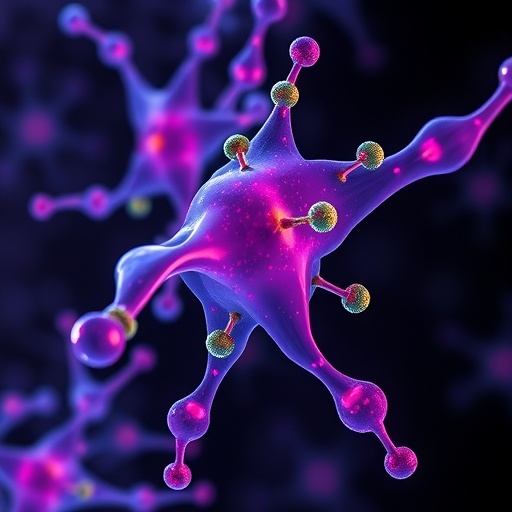
For decades, drug discovery has been dominated by a simple assumption: if a disease-driving protein cannot be reached by a conventional small molecule or an antibody administered outside the cell, it is effectively beyond therapeutic control. That assumption is now being challenged by a growing class of genetically encoded protein binders designed to operate inside living cells. In a review published in Nature Biomedical Engineering, Natalia V. Barykina and Vladislav V. Verkhusha describe how nanobodies, DARPins, affibodies and de novo-designed binding scaffolds are opening new routes to proteins that have remained inaccessible to traditional medicines. These compact molecules can recognize intracellular targets with high selectivity, report on their activity and, in some cases, redirect them toward destruction.
The need for such technologies is substantial. Many proteins implicated in cancer, neurodegeneration and infectious disease function in the cytoplasm, nucleus, mitochondria or other intracellular compartments, where extracellular antibodies cannot reach them unaided. Small molecules can enter cells, but they often bind to deep pockets that are absent from many regulatory proteins. Oncogenic transcription factors, scaffold proteins, protein–protein interaction surfaces and disease-associated conformations may present broad, shallow or highly flexible interfaces that are difficult for conventional chemistry to engage. Intracellular binders offer a different strategy. Rather than searching only for a drug-like pocket, researchers can engineer proteins with extended contact surfaces that wrap around a target or lock onto a particular structural state.
Nanobodies are among the best-known examples. Derived from the antigen-binding domains of camelid heavy-chain antibodies, these single-domain proteins are considerably smaller than conventional antibodies while retaining a defined antigen-recognition surface. Their compact architecture can improve access to crowded cellular environments and, in some cases, allow them to enter clefts or bind epitopes that are sterically inaccessible to larger molecules. DARPins, or designed ankyrin repeat proteins, use repeated structural modules to create stable, highly programmable binding surfaces. Affibodies are based on the small three-helix bundle of the staphylococcal protein A domain, while newer de novo scaffolds are built computationally or through protein engineering rather than adapted directly from natural antibodies. Each platform brings a different balance of size, stability, expression efficiency, specificity and ease of modification.
The genetic encoding of these binders is central to their versatility. Instead of delivering a purified protein, researchers can introduce DNA or RNA instructions that cause a cell to manufacture the binder internally. This approach makes it possible to place binders under tissue-specific, inducible or disease-responsive control. A binder can be fused to a fluorescent protein to visualize the location and movement of an endogenous target, linked to a degradation component to reduce the target’s abundance, or connected to a regulatory domain that changes the target’s activity. Because the binder is produced in the same cell as its target, it can function as a continuously present molecular probe rather than as a one-time reagent.
In imaging, these molecules are transforming the way scientists observe proteins in their native setting. A fluorescently tagged intracellular binder can recognize an endogenous protein without requiring the target to be genetically fused to a fluorescent marker. This distinction matters because large reporter tags can alter protein folding, trafficking or interactions. Binders can also be engineered to distinguish between active and inactive conformations, providing a visual readout of molecular state rather than simple protein abundance. In this way, a fluorescent binder may reveal where a signalling molecule becomes activated, how a pathogenic protein changes shape or when a regulatory complex assembles inside a living cell. Such tools are particularly valuable for studying dynamic processes that are invisible in fixed samples.
The same recognition principles can be converted into biosensors and control systems. A binder can be designed to change its fluorescence, localization or interaction partners when it captures a target. Coupled to engineered signalling modules, these responses can create intracellular circuits that detect disease-associated proteins and trigger a measurable output. In therapeutic research, binders have been used to inhibit oncogenic signalling by blocking interaction surfaces, sequester proteins in specific cellular compartments or interfere with the assembly of harmful complexes. Other designs act as molecular adaptors, bringing a target protein into proximity with the cell’s degradation machinery. These systems can reduce the amount of a disease-driving protein rather than merely suppressing its activity, a potentially important advantage when a target lacks a conventional inhibitory pocket.
Intracellular binders are also becoming components of programmable cell therapies. Engineered immune cells, for example, could use genetically encoded recognition modules to sense intracellular states indirectly through signalling pathways, or to control the abundance and localization of proteins that determine cellular behaviour. In cancer models, a binder aimed at an oncogenic factor could be expressed selectively in malignant cells, while in infectious disease research, binders may be designed to intercept pathogen proteins after infection. The ability to combine a recognition domain with degradation, imaging or signalling functions creates a modular design space in which one binder can serve as a sensor, inhibitor and therapeutic trigger, depending on its molecular attachments.
Finding effective intracellular binders, however, is not a straightforward matter of selecting the tightest interaction. Discovery platforms such as phage display, yeast display, ribosome or mRNA display and high-throughput screening can generate large libraries of candidate proteins, but candidates must then be tested under intracellular conditions. A molecule that binds strongly in a purified biochemical assay may misfold in the cytoplasm, aggregate, become trapped in the wrong compartment or fail to recognize the target in its native conformation. Researchers therefore evaluate not only affinity and specificity, but also folding, solubility, expression level, resistance to proteolysis and behaviour in the crowded cellular environment. The target itself may also be modified, partially unfolded or surrounded by competing partners, making cellular validation essential.
Computational protein design is beginning to expand this search space. Structure-based modelling can identify surfaces that may be suitable for binding and predict mutations that improve affinity or stability. Machine-learning methods can assist with sequence generation, structure prediction and the prioritization of candidates before experimental testing. In principle, computational systems could design binders against targets for which no natural antibody-like reagent exists, including transient conformations and previously neglected protein surfaces. Yet prediction remains only one part of the process. Intracellular performance depends on factors that are difficult to model precisely, including expression kinetics, subcellular localization, degradation rates and unintended interactions with the proteome. The most productive workflow is therefore likely to combine computational design with display technologies, structural analysis and direct testing in living cells.
The path from promising molecular tool to clinical therapy remains challenging. A binder must remain stable and functional over time, reach the correct cell type and compartment, avoid provoking toxic immune responses and be delivered with sufficient efficiency. Viral vectors can provide durable genetic expression, but they raise concerns involving manufacturing, tissue targeting, dose control and pre-existing immunity. Non-viral systems, including lipid nanoparticles and other nucleic-acid delivery technologies, may offer greater flexibility but must overcome barriers to tissue penetration and intracellular release. Immunogenicity is another concern, particularly for scaffolds derived from nonhuman proteins or containing sequences unfamiliar to the patient’s immune system. The review by Barykina and Verkhusha presents intracellular binders as a rapidly expanding bridge between molecular engineering, live-cell imaging and therapeutic design. Their ultimate impact will depend on whether researchers can convert impressive cellular demonstrations into precisely delivered, durable and safe medicines capable of controlling proteins once considered unreachable.
Cite Scienmag News
Ophelia Keating. (August 20, 2026). Intracellular Protein Binders Enable Imaging, Cellular Control, and Future Therapies. Scienmag. https://scienmag.com/intracellular-protein-binders-enable-imaging-cellular-control-and-future-therapies/
Ophelia Keating. "Intracellular Protein Binders Enable Imaging, Cellular Control, and Future Therapies." Scienmag, 20 August 2026, https://scienmag.com/intracellular-protein-binders-enable-imaging-cellular-control-and-future-therapies/. Accessed 6 September 2026.
Ophelia Keating. "Intracellular Protein Binders Enable Imaging, Cellular Control, and Future Therapies." Scienmag. August 20, 2026. https://scienmag.com/intracellular-protein-binders-enable-imaging-cellular-control-and-future-therapies/


