


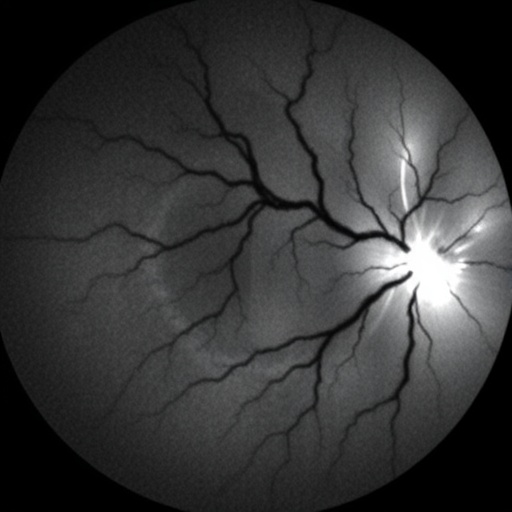
A new artificial intelligence system that reads the fine architecture of the human retina is drawing attention for its ability to trace even the thinnest blood vessels in fundus photographs, a task that has long frustrated standard deep learning pipelines. The system, called WRSG-Net, short for Wavelet-Refined Semantic-Guided Network, was developed by Hailu Liao, Guangle Yao, Zhanyong Mei and Sana Abbas at the College of Computer Science and Cyber Security, Chengdu University of Technology in Chengdu, China, and is described in an open access paper published in BMC Medical Imaging. Its central promise is straightforward but clinically consequential: more reliable computer-assisted detection of vascular damage in the eye, where diseases such as diabetic retinopathy and hypertensive retinopathy announce themselves first in tiny, easily missed vessels.
Retinal vessel segmentation, the pixel-by-pixel outlining of arteries and veins in an image of the eye’s fundus, is a foundational step in computer-aided diagnosis of a range of eye diseases. When a segmentation algorithm performs well, downstream software can quantify vessel tortuosity, detect narrowing, spot avascular zones and flag early signs of disease for ophthalmologists. When it performs poorly, the consequences ripple outward: thin capillaries vanish from the output map, vessel edges blur, and clinically meaningful detail is lost precisely where disease tends to appear first. The Chinese team argues that this is exactly the blind spot of existing methods. Fundus images pose a hostile environment for segmentation networks because vessels vary enormously in caliber, from thick arcades near the optic disc to capillaries barely a pixel wide; they branch into intricate, tree-like structures; they sit against backgrounds with low contrast; and they are frequently obscured by lesions or other pathological interference.
The dominant architectural template in medical image segmentation for years has been the encoder-decoder design, descended from the fully convolutional network and its famous cousin U-Net. In such designs, the encoder progressively downsamples the image, trading spatial resolution for increasingly abstract feature representations, and the decoder then upsamples those features back to full resolution to produce a segmentation mask. The trouble, as the authors explain, is that downsampling is ruthless with high-frequency information. Fine vessels, delicate branch points and sharp edges are encoded in the high-frequency components of an image, and repeated pooling operations can discard them irretrievably. What survives in the deepest layers tends to describe large structures well but thin structures badly, and decoder upsampling then compounds the problem by smearing whatever detail remains.
WRSG-Net attacks this problem from three directions, each embodied in a dedicated module. The first is a Parametric Wavelet Refined Encoder, or PWREC. Rather than relying solely on conventional downsampling, this encoder employs learnable wavelet decomposition, a mathematical technique adapted from signal processing in which an image is separated into components at different frequency bands and spatial locations. The decisive innovation is that the wavelet transform is parametric, meaning the network learns the decomposition itself during training rather than applying a fixed transform. This allows the encoder to adaptively capture high-frequency vascular detail, the fine texture and sharp transitions that characterize vessel boundaries, and to preserve it as the image passes through successive stages. The extracted features are then refined through a dual-attention mechanism, which learns to weight the most informative channels and spatial locations, mitigating the loss of fine vessel detail during downsampling.
The second component, the Inter-Scale Information Propagation Module, or ISIPM, addresses a different failure mode. In a typical encoder-decoder network, features at different scales are produced at different layers but often remain loosely coupled, joined only at skip connections that transfer them to the decoder. The ISIPM establishes an explicit layer-wise information propagation flow, allowing hierarchical features, from coarse global context down to fine local detail, to be integrated continuously across the network’s depth. The result the authors report is enhanced consistency and continuity in the segmented vessels: fewer breaks in the vascular tree, fewer disconnected fragments, and a more faithful reconstruction of the branching topology that clinicians rely on for assessment.
The third component is the Semantic Guided Decoder, or SGDC. Decoders commonly operate somewhat blindly, upsampling features with limited awareness of what the overall segmentation should look like. The SGDC instead performs context-aware feature aggregation under decoder semantic guidance, using high-level semantic information to direct how low-level features are assembled into the final pixel-level prediction. This design serves two purposes described by the authors: it suppresses noise originating in the encoder, preventing spurious activations from being painted into the output as phantom vessels, and it alleviates the blurring that typically accompanies decoder upsampling, sharpening vessel edges in the final mask.
Together, the three modules form a coherent design philosophy. Where prior approaches have often bolted attention blocks or multi-scale tricks onto otherwise conventional architectures, WRSG-Net reconsiders the information flow end to end, asking at each stage what detail is at risk of being lost and how it can be carried forward. The wavelet encoder protects high-frequency content at the point of greatest danger, downsampling; the inter-scale propagation module keeps hierarchical features talking to one another; and the semantic-guided decoder ensures that the final reconstruction is informed by high-level context rather than left to interpolate in the dark.
Benchmark performance is where such architectural claims must ultimately be judged, and the team evaluated WRSG-Net on the three most widely used public datasets for retinal vessel segmentation: DRIVE, STARE and CHASE_DB1. These datasets, each manually annotated by experts, serve as the de facto standard battlegrounds for segmentation algorithms, and their differing imaging conditions and patient populations make consistent performance across all three a meaningful achievement. On DRIVE, the Digital Retinal Images for Vessel Extraction dataset, WRSG-Net achieved an F1-score of 85.17 percent, accuracy of 97.84 percent, sensitivity of 84.31 percent and specificity of 97.88 percent. On STARE, the Structured Analysis of the Retina dataset, it reached an F1-score of 90.11 percent, accuracy of 98.04 percent, sensitivity of 85.06 percent and specificity of 98.71 percent. On CHASE_DB1, drawn from the Child Heart and Health Study in England, it recorded an F1-score of 84.11 percent, accuracy of 97.12 percent, sensitivity of 83.14 percent and specificity of 99.27 percent.
These numbers reward close reading. Accuracy and specificity are comparatively easy to inflate in vessel segmentation because the vast majority of pixels in a fundus image belong to the background; a network that simply predicted “no vessel” everywhere would score well above 90 percent accuracy. The metrics that separate strong systems from weak ones are sensitivity, the proportion of true vessel pixels correctly identified, and the F1-score, the harmonic mean of precision and recall that penalizes both missed vessels and false detections. WRSG-Net’s figures on all three benchmarks are competitive with, and on several measures superior to, most state-of-the-art approaches, according to the authors’ comparisons. More striking than the aggregate numbers, however, is where the gains concentrate. The team reports that the segmentation results show obvious advantages in the identification of tiny blood vessels and the repair of blurred edge details, which is precisely the regime where clinical value and technical difficulty both peak.
The clinical motivation behind the work is substantial. Diabetic retinopathy remains a leading cause of preventable blindness in working-age adults worldwide, and its earliest vascular signatures, microaneurysms, capillary dropout, venous beading, are subtle and require expert interpretation. Hypertensive retinopathy similarly encodes the history of blood pressure damage in the caliber and appearance of retinal vessels. Screening programs increasingly depend on automated image analysis to triage the enormous volumes of fundus photographs generated in population-scale programs, and the reliability of those programs is bounded by the fidelity of the underlying segmentation. A network that preserves capillary-level detail and clean vessel edges offers a stronger foundation for quantifying vascular change over time, and the authors position WRSG-Net as providing a new feasible technical path for retinal vessel segmentation with application potential in auxiliary screening and diagnosis of fundus vascular diseases.
The study was conducted entirely on publicly released, fully anonymized image datasets, involving no healthcare intervention on human participants, and the authors acknowledge the research teams behind the DRIVE, STARE and CHASE_DB1 benchmarks for their role in building the open resources on which the field depends. The research was supported by the Central Guidance on Local Science and Technology Development Fund Project (24ZYCXCP0020). The corresponding author is Guangle Yao of Chengdu University of Technology. The paper was received on 23 April 2026, accepted on 28 August 2026, and published on 05 September 2026 under open access terms that permit non-commercial sharing and reuse with attribution.
The broader significance of the work may lie in its demonstration that ideas from classical signal processing still have transformative power when fused with modern deep learning. Wavelets, which dominated image compression and denoising research for decades, fell out of fashion as convolutional networks learned their own feature hierarchies. WRSG-Net’s results suggest that the old and the new are complementary rather than competing: the wavelet transform offers an analytically sound way to represent multi-scale frequency content, while learned parameters and attention let the network decide which of those components matter for the task at hand. For the millions of patients whose eyes are photographed each year in screening programs, the difference between a segmentation that loses capillaries and one that keeps them may ultimately translate into earlier detection, better triage and preserved sight.
Cite Scienmag News
Ophelia Keating. (September 6, 2026). Wavelet-Enhanced Semantic Network Improves Retinal Vessel Segmentation Accuracy. Scienmag. https://scienmag.com/wavelet-enhanced-semantic-network-improves-retinal-vessel-segmentation-accuracy/
Ophelia Keating. "Wavelet-Enhanced Semantic Network Improves Retinal Vessel Segmentation Accuracy." Scienmag, 6 September 2026, https://scienmag.com/wavelet-enhanced-semantic-network-improves-retinal-vessel-segmentation-accuracy/. Accessed 6 September 2026.
Ophelia Keating. "Wavelet-Enhanced Semantic Network Improves Retinal Vessel Segmentation Accuracy." Scienmag. September 6, 2026. https://scienmag.com/wavelet-enhanced-semantic-network-improves-retinal-vessel-segmentation-accuracy/


