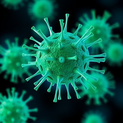
In the realm of biomedical science, the ability to identify and analyze microscopic cellular components is critical for advancing disease diagnosis and therapeutics. Among these components, exosomes—nanometer-sized extracellular vesicles secreted by most human cells—have emerged as pivotal players in intercellular communication, carrying molecular cargo that reflects the physiological state of their originating cells. Despite their profound potential, the detailed study of exosomes has been hampered by significant technological limitations, constraining our capacity to harness their full diagnostic and therapeutic promise. Recently, Wei Chuan Shih, a distinguished professor of electrical and computer engineering at the University of Houston, has pioneered a groundbreaking imaging technology that stands to revolutionize our understanding of these elusive biological nanoparticles.
The technology, bolstered by a $1.7 million grant from the National Institutes of Health, represents a leap forward in exosome analysis by enabling the examination of individual exosomes with unprecedented precision. Traditional analytical methods have struggled to achieve adequate sensitivity and specificity, often requiring large sample volumes and relying heavily on DNA amplification and sequencing techniques, which can obscure the nuanced heterogeneity among exosome populations. In contrast, Shih’s novel approach aims to surmount these challenges by directly measuring the structural and molecular attributes of single exosomes, thereby facilitating the identification of specific exosome subpopulations that may serve as viable drug targets.
Central to this breakthrough is the Integrated Nanophotonic Imaging and Spectroscopy Technology (INSPECT), a cutting-edge platform that leverages advances in nanoplasmonics to probe exosomes at a scale and depth previously unattainable. Nanoplasmonics, which exploits the interaction of light with metallic nanostructures to amplify electromagnetic fields, enables the detection of minute molecular binding events and enhances fluorescence signals. INSPECT uniquely combines three complementary nanoplasmonic mechanisms: surface binding detection, fluorescence signal enhancement, and chemical composition analysis, providing a multidimensional characterization of individual extracellular vesicles.
The biological significance of exosomes extends across numerous fields, including oncology, neurology, regenerative medicine, and dermatology. These vesicles mediate cell-to-cell communication by transporting proteins, lipids, and nucleic acids, influencing pathological processes such as tumor progression, neurodegeneration, and tissue repair. Harnessing exosomes for liquid biopsy applications—non-invasive sampling of biomarkers from bodily fluids—and drug delivery systems holds transformational potential for precision medicine. Nonetheless, the heterogeneity and complexity of exosomes present formidable obstacles to their clinical translation.
Current techniques suffer from multiple pitfalls: they often require extensive sample preparation steps like purification, isolation, and labeling, which can introduce artifacts and impede high-throughput analysis. Isolation procedures can be time-consuming and may result in the loss of critical subpopulations of vesicles. Furthermore, existing tools lack the resolution to perform multi-parametric profiling at the single-exosome level, meaning that subtle but biologically significant variations remain undetected. INSPECT’s capacity to integrate structural imaging with spectral analysis addresses these shortcomings by enabling simultaneous assessments of size, morphology, molecular composition, and binding interactions on a per-exosome basis.
Shih’s prior research laid the foundation for this innovation by demonstrating the efficacy of three nanoplasmonic enhancing modalities for biosensing. First, plasmonic resonance sensors detect molecular binding events via changes in the refractive index near metallic surfaces, providing sensitive label-free detection. Second, plasmon-enhanced fluorescence amplifies signal intensity, improving detection limits without increasing background noise. Third, surface-enhanced Raman scattering (SERS) offers detailed molecular “fingerprinting” by amplifying vibrational spectra, revealing chemical compositions with high specificity. The integration of these modalities within INSPECT creates a synergistic platform capable of extracting rich, multidimensional data from single exosomes.
The profound implications of this technology extend beyond fundamental science into translational and clinical realms. By enabling multiplexed, high-throughput analysis of exosome populations, INSPECT has the potential to identify novel biomarkers indicative of disease states, monitor therapeutic responses, and facilitate the development of exosome-based drug delivery vehicles. Moreover, this approach could accelerate research into the roles of exosomes in neurological disorders such as Alzheimer’s disease, where early and accurate detection of pathological changes is paramount.
Despite the promise, challenges remain in scaling the technology for widespread adoption. The sensitivity and specificity of INSPECT must be rigorously validated across diverse biological samples and conditions. Collaborations with biologists, clinicians, and other engineers are crucial to tailor the platform for various applications, optimize sample processing protocols, and ensure compatibility with existing diagnostic workflows. Shih emphasizes the collaborative nature of this endeavor, inviting researchers interested in extracellular vesicle biology to partner in expanding the capabilities and utility of INSPECT.
In sum, the advent of Integrated Nanophotonic Imaging and Spectroscopy Technology marks a significant milestone in the study and application of exosomes. By illuminating these diminutive yet biologically potent vesicles with unprecedented clarity, this innovation opens vistas for novel diagnostic and therapeutic strategies that could transform patient care across oncology, neurology, and beyond. As the scientific community embraces and refines such technologies, the once opaque world of exosomes will become increasingly transparent, revealing new molecular signatures and intervention points that hold the key to combating some of the most challenging diseases of our time.
Subject of Research: Exosomes; Integrated nanophotonic imaging and spectroscopy technology for single exosome analysis; biomedical engineering.
Article Title: Illuminating the Invisible: Nanophotonic Advances in Single Exosome Analysis Unveil New Paths for Disease Diagnosis and Treatment.
News Publication Date: Not specified.
Web References: University of Houston (https://uh.edu), National Institutes of Health (https://nih.gov).
Image Credits: University of Houston.
Keywords: exosomes, nanoplasmonics, integrated nanophotonic imaging, spectroscopy technology, single exosome analysis, biomedical engineering, disease diagnostics, drug delivery, extracellular vesicles, liquid biopsy, Alzheimer’s disease, cancer biology, nanomedicine, biomedical technology.
