


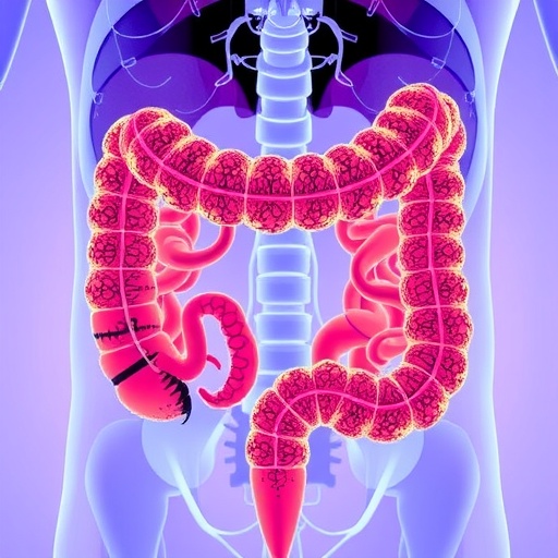
In a groundbreaking advancement at the intersection of oncology and artificial intelligence, researchers from the Faculty of Information Technology at the University of Jyväskylä have unveiled an AI-powered methodology that significantly enhances the analysis of colorectal cancer tissue samples. This innovative approach not only accelerates the diagnostic process but also offers precise predictions concerning the functionality of the DNA repair mechanisms within cancer cells, notably the mismatch repair (MMR) system. The implications of this research are vast, bearing potential to transform clinical workflows, reduce costs, and improve patient outcomes in colorectal cancer treatment.
Colorectal cancer, a leading cause of cancer-related morbidity worldwide, often demands laborious diagnostic procedures involving meticulous examination of tissue biopsies by pathologists. Central to the malignancy’s prognosis and treatment strategy is the assessment of the MMR system, a cellular mechanism responsible for correcting replication errors in DNA. Dysfunction in this error-correcting system contributes to tumor development and influences therapeutic decisions. Traditionally, evaluating the MMR status relies heavily on time-intensive manual analysis, a bottleneck that this new AI paradigm aims to dissolve.
The research, spearheaded by Liisa Petäinen and her team at the University of Jyväskylä, demonstrates that AI can shorten the turnaround time for such analyses from several days to mere minutes, without compromising accuracy. This acceleration not only expedites diagnosis but also implies potential cost reductions and more efficient utilization of scarce pathology expertise, enabling healthcare providers to reallocate specialist resources to other critical areas.
Unlike conventional techniques that concentrate exclusively on tumor zones at high magnification—typically a twentyfold zoom—the new AI framework exploits lower magnification levels, such as fivefold, to scan broader tissue regions encompassing both tumor and adjacent non-tumor areas. Intriguingly, the results underscore that peritumoral tissue harbors informative features related to DNA repair function, suggesting that a holistic tissue analysis can improve diagnostic precision. This holistic assessment could one day render the pre-identification of tumor regions unnecessary, streamlining diagnostic pipelines further.
The AI model, rigorously trained on a comprehensive dataset derived from approximately 1,300 colorectal cancer patient samples furnished by the Central Finland Biobank, has exhibited robustness beyond its original environment. Validated with external data sourced from Oulu University Hospital and American institutions, the model demonstrated high reliability across diverse populations and clinical settings, underscoring the potential for widespread applicability.
Finland’s unique healthcare infrastructure, characterized by integrated biobanking, unified patient registries, and interoperable clinical systems, has been pivotal to this research’s success. Tiina Jokela, Research Director at the Wellbeing Services County of Central Finland, affirms that this synergy between clinical practice and cutting-edge AI research fosters rapid translation from innovation to real-world healthcare solutions. The collaborative milieu of Central Finland, supplemented by regional funding and European Union co-financing under the AI-Hub II project umbrella, exemplifies how strategic partnerships and resource alignment accelerate scientific breakthroughs.
Moreover, the AI model leverages prior developments from Central Finland’s earlier projects, collectively improving cancer detection algorithms and refining predictive capabilities related to DNA mismatch repair deficiencies. This iterative enhancement process, grounded in large-scale datasets encompassing longitudinal patient histories from the years 2000 to 2015, enables the AI to recognize complex histopathological signatures that may elude conventional microscopic analysis.
Pathologists stand to benefit considerably from this technology. By automating routine evaluations, AI releases valuable human resources to focus on more nuanced diagnostic challenges and research. This interplay between human expertise and machine precision epitomizes the future of pathological diagnosis, where AI serves as an augmentative tool rather than a replacement, enhancing both efficiency and accuracy.
Critically, the study emphasizes that while early results are promising, thorough validation with larger, multi-institutional datasets remains essential. This ensures the AI’s generalizability, reproducibility, and safety before routine clinical deployment. It also highlights the necessity for continued collaboration among data scientists, clinicians, and regulatory bodies to address challenges related to ethics, patient privacy, and data security.
From a technical standpoint, the AI protocol integrates complex machine learning architectures capable of parsing intricate tissue morphology patterns at varying magnifications. By applying convolutional neural networks trained on annotated histopathological images, the system quantifies morphological and textural features linked to the malfunctioning of the MMR pathway. Through this, it offers a probabilistic assessment of repair mechanism status, which is critical for tailoring immunotherapy approaches and DNA-damage response-targeted treatments.
This transformative research not only paves the way for faster and more accurate colorectal cancer diagnostics but also sets a precedent for integrating AI into oncological histopathology more broadly. As digital pathology infrastructure grows globally, such AI-driven tools bear the promise to democratize access to expert-level diagnostics, particularly in resource-constrained healthcare environments.
In summary, the University of Jyväskylä and its partners have leveraged Finland’s rich biomedical data assets and cutting-edge AI methodologies to create an innovative diagnostic tool that accelerates colorectal cancer tissue analysis while providing precise insights into critical DNA repair processes. This fusion of technology and medicine heralds a new era of personalized, efficient cancer care with profound implications for patients, clinicians, and healthcare systems worldwide.
News Publication Date: 17-Mar-2026
Web References:
DOI: 10.1016/j.cmpb.2026.109317
Cite Scienmag News
Nathaniel Bowman. (May 28, 2026). Innovative AI Model Enhances Efficiency in Analyzing Colorectal Cancer Samples. Scienmag. https://scienmag.com/innovative-ai-model-enhances-efficiency-in-analyzing-colorectal-cancer-samples/
Nathaniel Bowman. "Innovative AI Model Enhances Efficiency in Analyzing Colorectal Cancer Samples." Scienmag, 28 May 2026, https://scienmag.com/innovative-ai-model-enhances-efficiency-in-analyzing-colorectal-cancer-samples/. Accessed 4 September 2026.
Nathaniel Bowman. "Innovative AI Model Enhances Efficiency in Analyzing Colorectal Cancer Samples." Scienmag. May 28, 2026. https://scienmag.com/innovative-ai-model-enhances-efficiency-in-analyzing-colorectal-cancer-samples/


