


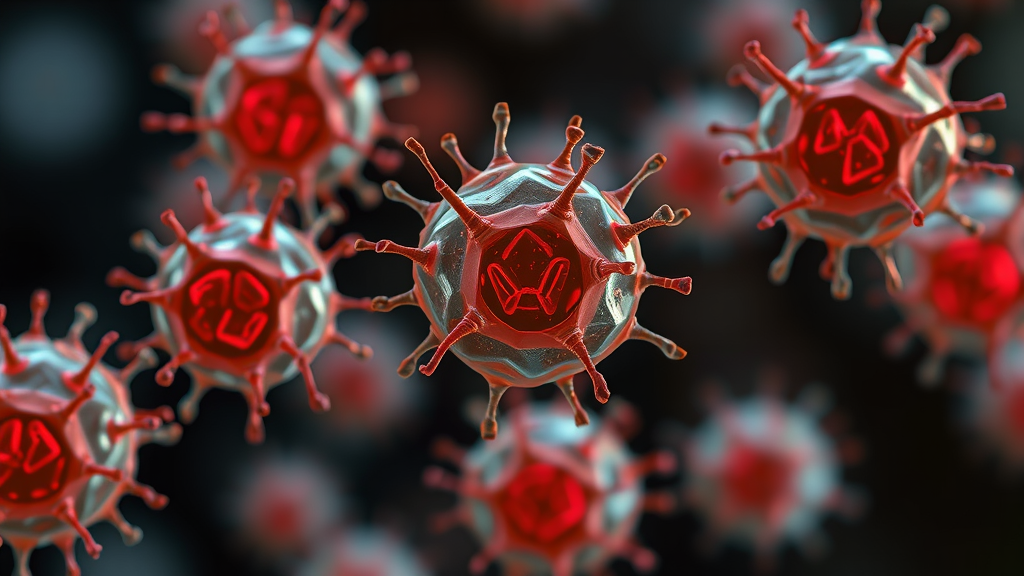
A viral infection that never reaches the egg itself can still derail its development, according to new research from Hokkaido University that reveals a surprising route by which viruses in the female reproductive tract may undermine fertility. The study, published in Biochemical Genetics, shows that when cumulus cells—the specialized support cells surrounding a freshly ovulated egg—are infected by an RNA virus, they mount a vigorous antiviral response whose inflammatory signaling molecules leak into the egg’s immediate environment and impair its ability to develop into a healthy embryo. The work, conducted by Keisuke Sasaki and Manabu Kawahara of the Laboratory of Animal Genetics and Reproduction at Hokkaido University’s Research Faculty of Agriculture, offers the clearest evidence to date that the maternal inflammatory microenvironment, rather than direct viral invasion, can be the decisive factor in early reproductive failure.
The research team set out to address a persistent gap in reproductive virology. Viral infections in the female reproductive tract are well known to pose risks to fertility, and previous studies in mice have shown that viral infection of the ovaries can compromise pregnancy. Yet the specific impact of viruses on ovulated oocytes—the mature eggs that have just been released from the ovary—and the role played by the surrounding cumulus cells remained poorly understood. Cumulus cells form a layered, cloud-like structure called the cumulus oophorus around the oocyte, and together the egg and its companion cells are known as the cumulus–oocyte complex, or COC. This intimate relationship is metabolically essential: the oocyte depends on cumulus cells for nutrients, signaling molecules, and developmental cues throughout its maturation. What Sasaki and Kawahara wanted to know was whether this dependency could become a liability during a viral attack.
To model the situation, the researchers used vesicular stomatitis virus, or VSV, a bullet-shaped RNA virus that is a standard laboratory tool for studying antiviral immunity. VSV enters cells through clathrin-dependent endocytosis and replicates rapidly in the cytoplasm, making it a reliable trigger of the innate immune pathways that cells use to detect RNA viruses. The team exposed mouse cumulus–oocyte complexes to the virus and then assessed how the cells responded at the level of gene expression, using quantitative real-time PCR to measure antiviral transcripts. In parallel, they tracked developmental outcomes by fertilizing the exposed oocytes in vitro and counting how many progressed through cleavage divisions and on to the blastocyst stage, the last step before implantation.
The gene expression analysis revealed a striking asymmetry between the two cell types in the complex. Ovulated oocytes did express retinoic acid-inducible gene-I, known as RIG-I, which is the cytosolic receptor that detects RNA viruses inside infected cells. But the oocytes lacked expression of two other key sensors of the RIG-I family: melanoma differentiation-associated gene 5, or MDA5, and laboratory of genetics and physiology 2, or LGP2. These helicase genes were present in the cumulus cells. The finding matters because the RIG-I family of DExD/H-box helicases forms the front line of intracellular RNA virus detection, with RIG-I and MDA5 recognizing different classes of viral RNA and LGP2 acting as a regulatory partner that fine-tunes their activity. The differential expression suggests that the oocyte’s antiviral surveillance toolkit is incomplete, and that its defenses may rely heavily on the completeness of the cumulus cells’ immune machinery.
When intact cumulus–oocyte complexes were exposed to VSV, the consequences for development were clear. The virus significantly impaired preimplantation development, reducing both the rate at which fertilized eggs underwent cleavage and the rate at which embryos formed blastocysts. Yet when the researchers looked for evidence of actual viral infection inside the oocytes and early embryos, they found none. The authors attribute this protection to the zona pellucida, the glycoprotein shell that surrounds the oocyte and early embryo and acts as a physical barrier. This result reframes the problem: the damage to development occurs without the virus ever setting foot inside the cell it ultimately harms.
Several follow-up experiments cemented the indirect mechanism. First, when the researchers stripped the cumulus cells away and exposed denuded oocytes directly to VSV, the oocytes neither induced antiviral gene expression nor showed developmental defects. On their own, the eggs simply did not respond to the virus. Second, and most tellingly, when uninfected oocytes were co-cultured with VSV-infected cumulus cells, their development was impaired—demonstrating that the mere presence of infected neighbors, with no virus reaching the oocyte, was sufficient to cause the damage. The virus, in effect, converted the egg’s own nurse cells into a source of developmental toxicity.
The molecular signature of the infected cumulus cells explained why. The infected cells exhibited a robust antiviral response, with significant upregulation of RIG-I itself, interferon-beta, interleukin-6, and tumor necrosis factor-alpha. Interferon-beta is the classic first-responder signal of the antiviral state, while interleukin-6 and tumor necrosis factor-alpha are inflammatory cytokines that can act on neighboring cells. Crucially, the researchers found that oocytes and zygotes express the receptor subunits for interleukin-6, encoded by the genes Il6ra and Gp130. This means the egg is structurally equipped to receive and respond to IL-6 signals arriving from its surroundings. The interleukin-6 pathway is already known to play roles in preimplantation embryos, where the IL-6 family cytokine leukemia inhibitory factor is essential for implantation, and the IL-6/STAT3 axis has been linked to anti-apoptotic signaling in mouse embryos. The new data identify IL-6 as a candidate mediator of the developmental impairment caused by infected cumulus cells.
The study’s authors frame the findings as revealing both the protective and the vulnerable nature of the cumulus–oocyte complex during viral challenge. The cumulus cells act as a shield: their complete antiviral sensor repertoire allows them to detect and respond to the virus, and the physical barrier of the zona pellucida keeps the virus out of the oocyte. But the same activation that defends the complex also floods the perivitelline environment with inflammatory cytokines, and the oocyte, which lacks its own full complement of viral sensors, appears susceptible to the paracrine consequences. The work thus provides a mechanistic account of how the maternal inflammatory microenvironment can influence early embryonic success, even in the absence of direct infection of the embryo itself.
The implications extend to a broader literature on viral infection and fertility. Hepatitis E virus has been shown to replicate in the ovary and promote oocyte apoptosis in rabbits, and Zika virus has been shown to cause acute infection and inflammation in the mouse ovary, with sexual transmission routes documented in mouse models. Herpes simplex virus type 2 sheds asymptomatically in the human female genital tract, and viral infection of the ovaries has been shown to compromise pregnancy while also revealing innate immune mechanisms that protect fertility. The new study adds a distinct mechanism to this list: not direct ovarian infection, and not viral tropism for the gamete, but the transformation of the egg’s own supporting cells into cytokine factories that compromise its developmental competence. This pathway could be relevant to unexplained fertility deficits associated with systemic or reproductive tract viral illness.
For the assisted reproduction field, the results suggest that the health of cumulus cells is not merely a marker of oocyte quality but an active determinant of embryo outcomes under immune challenge. The work was supported by JSPS KAKENHI grants 24K09199, awarded to Sasaki, and 24K01902, awarded to Kawahara, and all animal experiments were approved by the Regulatory Committee for the Care and Use of Animals of Hokkaido University. The authors note that the datasets supporting the developmental rate findings are available in the supplementary materials, with other data available from the corresponding author on reasonable request. Future work, the study implies, will need to test whether blocking interleukin-6 signaling during viral illness can rescue the developmental potential of exposed oocytes, and whether the same paracrine mechanism operates in other species, including humans—questions that could shape how fertility preservation is approached in patients confronting acute viral infections of the reproductive tract.
Subject of Research: Antiviral responses of mouse cumulus–oocyte complexes and indirect viral impairment of oocyte developmental competence via cumulus cell inflammatory signaling.
Article Title: Viral Infection of Cumulus Cells Impairs the Developmental Competence of Ovulated Mouse Oocytes
Article References: Sasaki, K., & Kawahara, M. (2026). Viral Infection of Cumulus Cells Impairs the Developmental Competence of Ovulated Mouse Oocytes. Biochemical Genetics. https://doi.org/10.1007/s10528-026-11449-4
Image Credits: AI Generated
DOI: 10.1007/s10528-026-11449-4
Keywords: oocyte, cumulus cells, vesicular stomatitis virus, antiviral response, RIG-I, interleukin-6, zona pellucida, fertility, preimplantation embryo, paracrine signaling, viral infection, reproductive immunology
Cite Scienmag News
Kristina Jarvis. (September 20, 2026). Infected Nurse Cells Sabotage Egg Development Through Inflammatory Signals. Scienmag. https://scienmag.com/infected-nurse-cells-sabotage-egg-development-through-inflammatory-signals/
Kristina Jarvis. "Infected Nurse Cells Sabotage Egg Development Through Inflammatory Signals." Scienmag, 20 September 2026, https://scienmag.com/infected-nurse-cells-sabotage-egg-development-through-inflammatory-signals/. Accessed 4 October 2026.
Kristina Jarvis. "Infected Nurse Cells Sabotage Egg Development Through Inflammatory Signals." Scienmag. September 20, 2026. https://scienmag.com/infected-nurse-cells-sabotage-egg-development-through-inflammatory-signals/


