


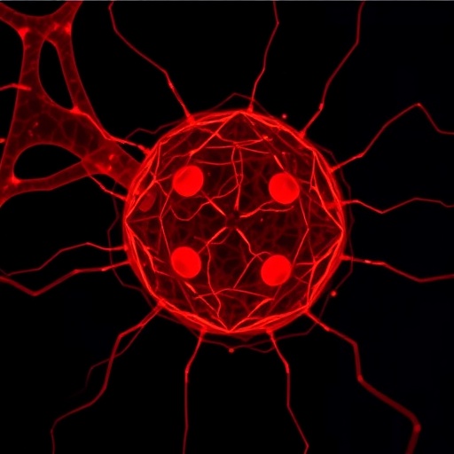
In a remarkable leap forward for cellular biology and nanotechnology, researchers have unveiled a pioneering technique to visualize single extracellular vesicles (EVs) with unprecedented clarity. These minuscule biological structures, which carry critical molecular messages across cells, have eluded high-resolution analysis due to their nanoscale size and heterogeneity. The new method combines rolling circle amplification with expansion microscopy, offering a groundbreaking approach that could radically enhance our understanding of intercellular communication and pathogenesis.
Extracellular vesicles, ranging from exosomes to microvesicles, serve as vital couriers packed with proteins, lipids, and nucleic acids, mediating physiological and pathological processes. Despite their importance, direct imaging at the single-vesicle level has been notoriously challenging. Conventional fluorescence microscopy is limited by the diffraction barrier, while electron microscopy, although high in resolution, lacks molecular specificity and throughput. Ultra-sensitive techniques capable of pinpointing both structural and biomolecular details at the nanoscale are therefore highly sought after.
The research team, led by Wu, J., Dou, Q., and Mao, M., has ingeniously integrated rolling circle amplification (RCA) with expansion microscopy (ExM) to circumvent these limitations. RCA is a nucleic acid amplification process that generates long, single-stranded DNA concatemer strands from circular templates, which in this context enables amplification of molecular signals tethered to the EVs. Expansion microscopy physically enlarges the biological specimen through a swellable polymer, effectively magnifying nanoscale features into the microscale range compatible with standard optical microscopy.
This dual-strategy method markedly enhances fluorescence signal intensity while spatially separating densely packed molecules inside the vesicles. Specifically, the team first labeled EV-associated molecules with DNA probes designed to initiate RCA. As RCA proceeds, the amplified DNA products yield bright fluorescent signals, which are then spatially expanded by the hydrogel-based ExM process. This systematic expansion magnifies the vesicles roughly fourfold in linear dimension, translating to over 60-fold volumetric enlargement, allowing for detailed imaging via confocal microscopes that are widely available.
One of the key breakthroughs lies in the protocol’s sensitivity and specificity for single extracellular vesicles, which traditionally blend into complex biological milieus and are often mischaracterized en masse. This refined visualization enables differentiation among vesicle subpopulations based on molecular cargo and size, revealing heterogeneity that is critical for both fundamental biology and clinical diagnostics. The ability to track individual vesicles may elucidate their roles in disease progression and therapeutic delivery mechanisms.
Furthermore, the RCA–ExM technique holds promise for multiplexed analysis, as various molecular targets can be amplified and visualized simultaneously by using distinct rolling circle amplification probes and fluorophores. This multiplexing is crucial for mapping the diverse molecular landscapes of EVs, which harbor distinct biomolecular signatures depending on cellular origin and pathological conditions such as cancer, neurodegeneration, or viral infections.
In practical applications, this method promises to revolutionize liquid biopsy approaches by enabling high-resolution profiling of circulating extracellular vesicles from patient blood samples. Such detailed molecular readouts could refine early disease detection, monitor treatment efficacy, and unravel mechanisms of metastasis or immune modulation. This is a pivotal shift from bulk analyses towards precision diagnostics at the single vesicle level.
Beyond medical diagnostics, the technique sets a new benchmark for fundamental research, potentially reshaping our understanding of vesicle biology in neuroscience, immunology, and developmental biology. Researchers can now probe vesicle biogenesis, cargo sorting, and cell-to-cell communication dynamics with unprecedented granularity, providing insights that were previously unattainable.
The RCA–ExM approach also boasts compatibility with existing laboratory equipment, democratizing access to super-resolved EV imaging. This lowers the barrier to adoption and accelerates translational research efforts. Its utilization of standard fluorescence microscopy platforms means that laboratories across the globe can implement this technique without the need for costly specialized instruments.
Intriguingly, the physical expansion aspect of the method does not significantly distort molecular spatial relationships, preserving the biophysical context necessary for accurate interpretation. This ensures that researchers can interpret vesicle architecture and cargo distribution with confidence, avoiding artifacts that have plagued other amplification or labeling strategies.
Moreover, the method is robust and adaptable to a wide range of biological samples, enabling the study of EVs derived from complex tissue matrices, bodily fluids, and cultured cells. This versatility widens its impact, facilitating cross-disciplinary research that spans from clinical pathology to nanoscale biophysics.
The study’s findings pave the way for future innovations that could integrate RCA–ExM with live-cell imaging or single-molecule tracking, further enhancing temporal and spatial resolution. Such advancements would bridge the gap between static snapshots and dynamic processes, uncovering the real-time behavior of extracellular vesicles in vivo.
This work also contributes to the broader scientific endeavor of super-resolution imaging, joining a suite of emerging techniques that challenge the limits of optical microscopy. By merging molecular amplification with physical expansion, it delivers a cost-effective and scalable route to nanoscale resolution, which is an exciting paradigm for various subcellular imaging challenges beyond EVs.
As extracellular vesicles continue to gain significance as biomarkers and therapeutic vehicles, technologies like the one developed by Wu, Dou, Mao, and colleagues will be instrumental in validating their clinical utility. Precise visualization and molecular profiling are critical steps to translate EV research from bench to bedside, and this method surmounts longstanding technical barriers.
In summary, the integration of rolling circle amplification with expansion microscopy represents a transformative advance, enabling single extracellular vesicle imaging with remarkable sensitivity and resolution. The technique broadens the horizons of EV research, offering a window into the previously inaccessible nanoworld of intercellular communication and molecular diagnostics. Its adoption could spur new discoveries across biology and medicine, redefining our capability to explore cellular microcosms.
Cite Scienmag News
Ophelia Keating. (August 13, 2025). Imaging Single Extracellular Vesicles with RCA-Expansion. Scienmag. https://scienmag.com/imaging-single-extracellular-vesicles-with-rca-expansion/
Ophelia Keating. "Imaging Single Extracellular Vesicles with RCA-Expansion." Scienmag, 13 August 2025, https://scienmag.com/imaging-single-extracellular-vesicles-with-rca-expansion/. Accessed 4 September 2026.
Ophelia Keating. "Imaging Single Extracellular Vesicles with RCA-Expansion." Scienmag. August 13, 2025. https://scienmag.com/imaging-single-extracellular-vesicles-with-rca-expansion/


