


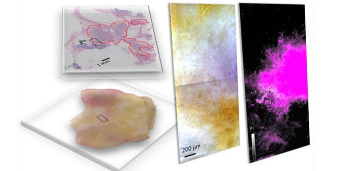
Breast-conserving surgery (BCS), also called lumpectomy, involves the removal of a cancerous lump and some surrounding tissue. BCS is suitable for women with early-stage breast cancer or small lumps, as it preserves more of the breast compared to mastectomy. After BCS, it is crucial to ensure that all cancer cells are removed to determine if further surgery is needed. This is done through a tumor margin assessment, where the edges of the removed area (tumor margins) are examined for the presence of cancer cells. Typically, tumor margin assessments involve staining tissue samples with dyes and examining them under a microscope to distinguish healthy cells from cancer cells. However, emerging optical imaging methods offer faster alternatives for this assessment.

Credit: Image courtesy of Jeeseong Hwang from the National Institute of Standards and Technology.
Breast-conserving surgery (BCS), also called lumpectomy, involves the removal of a cancerous lump and some surrounding tissue. BCS is suitable for women with early-stage breast cancer or small lumps, as it preserves more of the breast compared to mastectomy. After BCS, it is crucial to ensure that all cancer cells are removed to determine if further surgery is needed. This is done through a tumor margin assessment, where the edges of the removed area (tumor margins) are examined for the presence of cancer cells. Typically, tumor margin assessments involve staining tissue samples with dyes and examining them under a microscope to distinguish healthy cells from cancer cells. However, emerging optical imaging methods offer faster alternatives for this assessment.
In a study published in the Journal of Biomedical Optics, researchers from the United States introduced hyperspectral dark-field microscopy (HSDFM) as a method to rapidly and accurately differentiate between cancerous and healthy cells, as well as to identify different tumor subtypes within breast tissues following lumpectomy procedures. “We successfully identified specific regions containing carcinoma subtypes, including invasive ductal carcinoma and invasive mucinous carcinoma, in freshly excited tissues by applying machine learning algorithms to the imaging data,” author Jeeseong Hwang explains.
In HSDFM, tissue samples are illuminated by multiple wavelengths of light, and the differences in the intensity of scattered light as a function of wavelength from cellular and molecular substances are analyzed to produce unique spectral signatures of the tissues. The method produces a two-dimensional image where each pixel contains spectral information across multiple wavelengths, making it capable of identifying the composition of tissues. This imaging method specifically addresses issues persistent in most hyperspectral tumor margin imaging techniques, which rely on reflectance to gather spectral data of tissue samples. Reflectance-based methods encounter challenges due to the uneven absorption of light by biological substances such as oxyhemoglobin in blood, leading to inconsistent spectral signatures across multiple samples.
In this study, the researchers analyzed HSDFM images of breast lumpectomy samples taken from multiple patients. To classify the pixels according to the tissue type, they used two different approaches: supervised and unsupervised machine learning.
For the supervised approach, the researchers utilized a method called spectral angle mapping, which compares the spectral signature of each pixel in the hyperspectral image to known spectral signatures of tumor subtypes and tissue types (such as fat, interconnected tissue, and blood) identified through histopathological analysis. For the unsupervised approach, they utilized the K-means clustering algorithm. This method groups pixels with similar spectral signatures into clusters, facilitating the identification of tumor regions without prior knowledge of reference spectra or tissue types.
The spectral signatures obtained by both methods were quite similar and effectively pinpointed regions with invasive ductal carcinoma, the most common type of breast cancer, representing 75 percent of all breast cancer cases, as well as invasive mucinous carcinoma, a rare type characterized by cancer cells developing in mucus.
Breast cancer is a leading cause of cancer-related deaths in women. This study shows that the unsupervised method is validated by the supervised method, therefore HSDFM imaging data can be used to develop unsupervised algorithms for the rapid and accurate identification of cancerous tissues. Thus, it is expected to enhance postsurgical care for BCS and facilitate timely corrective actions.
For details, see the Gold Open Access article by J. Hwang et al., “Hyperspectral dark-field microscopy of human breast lumpectomy samples for tumor margin detection in breast-conserving surgery,” J. Biomed. Opt. 29(9), 093503 (2024), doi 10.1117/1.JBO.29.9.093503.
Journal
Journal of Biomedical Optics
Article Title
Hyperspectral dark-field microscopy of human breast lumpectomy samples for tumor margin detection in breast-conserving surgery
Article Publication Date
7-May-2024
Cite Scienmag News
Drew Townsend. (May 9, 2024). Hyperspectral dark-field microscopy for rapid and accurate identification of cancerous tissues. Scienmag. https://scienmag.com/hyperspectral-dark-field-microscopy-for-rapid-and-accurate-identification-of-cancerous-tissues/
Drew Townsend. "Hyperspectral dark-field microscopy for rapid and accurate identification of cancerous tissues." Scienmag, 9 May 2024, https://scienmag.com/hyperspectral-dark-field-microscopy-for-rapid-and-accurate-identification-of-cancerous-tissues/. Accessed 5 September 2026.
Drew Townsend. "Hyperspectral dark-field microscopy for rapid and accurate identification of cancerous tissues." Scienmag. May 9, 2024. https://scienmag.com/hyperspectral-dark-field-microscopy-for-rapid-and-accurate-identification-of-cancerous-tissues/

