


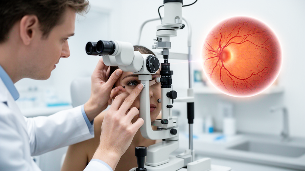
The human retina has long been described as a window into the brain, and a new analysis of tens of thousands of adults suggests that window is clearer than ever. In a large study drawing on the UK Biobank, researchers report that people who score higher on a composite measure of brain-healthy habits—the brain care score—tend to have measurably thicker retinal nerve layers, thicker photoreceptor layers, and a retina that appears biologically younger than their chronological age. The work, published in GeroScience, is the first to connect this particular brain health index to detailed structural measurements of the eye, and it offers a strikingly concrete way to see the consequences of everyday health choices.
The brain care score, developed at the McCance Center for Brain Health, condenses twelve modifiable factors into a single number: four physical measures (blood pressure, hemoglobin A1c, cholesterol, and body mass index), five lifestyle behaviors (nutrition, alcohol intake, smoking, physical activity, and sleep), and three social-emotional dimensions (stress, relationships, and meaning in life). In the UK Biobank adaptation used here, the score ranges up to 19 points, with higher values indicating better brain care. Previous studies have linked higher scores to favorable neuroimaging markers and reduced risks of dementia, stroke, and late-life depression. What remained unknown was whether the score also tracks with the fine-grained architecture of the retina, an outgrowth of the central nervous system that shares vascular, metabolic, and inflammatory biology with the brain.
To find out, the team led by investigators at The Chinese University of Hong Kong analyzed optical coherence tomography (OCT) scans from 28,657 UK Biobank participants aged 40 to 69. OCT is a non-invasive imaging technique that uses light interference to map retinal layers at micrometer resolution, and the researchers examined eight distinct layers plus overall macular thickness. After rigorous quality control and statistical adjustment for age, sex, ethnicity, education, deprivation, cardiovascular disease history, refractive error, and intraocular pressure, a clear pattern emerged: each 5-point increase in the brain care score was associated with thicker retinal nerve fiber layer, thicker ganglion cell–inner plexiform layer, thicker photoreceptor segments, and greater average macular thickness.
The effect sizes are small in absolute terms—a few tenths of a micrometer per 5-point score increase, translating to roughly 1.2 percent difference in nerve fiber layer thickness and about half a percent for the ganglion cell layer relative to cohort means. The researchers are careful to stress that these are population-level structural differences, not differences an ophthalmologist could detect in a single patient or act on clinically. Yet the pattern was remarkably consistent. Participants in the lowest score quartile had significantly thinner layers across the board compared with those in the highest quartile, all trend tests were highly significant, and the findings held up in sensitivity analyses that accounted for nonlinear aging effects, restricted the sample to European participants, and replicated the associations using repeat OCT visits years later.
Perhaps the most provocative result concerns retinal biological age. The team trained a support vector machine on 60 OCT-derived structural metrics from thousands of healthy participants to predict chronological age from retinal structure alone, then defined an OCT age gap as the bias-corrected predicted age minus actual age. Positive values indicate a retina that looks older than it should. After full adjustment, each 5-point increase in brain care score was associated with a 0.326-year reduction in this retinal age gap, with the mean gap falling progressively from 0.39 years in the lowest score quartile to −0.08 years in the highest. In other words, people who take better care of their brains carry retinas that read as younger on a machine-learning clock.
Dissecting the score into its components revealed where the signal lives. The physical and lifestyle domains drove the associations, while the social-emotional domain showed no independent link to retinal structure. Favorable blood pressure, hemoglobin A1c, and body mass index were each tied to thicker specific layers and a smaller retinal age gap, with effects on the age gap ranging from about a quarter to nearly half a year. Non-smoking, moderate alcohol intake, regular aerobic exercise, and at least seven hours of nightly sleep each contributed modest reductions in retinal biological age. Interestingly, total cholesterol below 190 milligrams per deciliter was actually associated with a slightly larger retinal age gap, echoing a growing literature on the complex, sometimes U-shaped relationships between lipids and neurological health.
To probe the biology behind these correlations, the researchers turned to metabolomics. For a subset of 14,656 participants with nuclear magnetic resonance profiling of 249 plasma biomarkers, they ran exploratory mediation analyses asking whether circulating metabolites statistically carry part of the association between brain care score and retinal thickness. The candidate pathways that emerged centered on lipid metabolism, unsaturated fatty acids, and branched-chain amino acids. One principal component reflecting HDL-enriched lipid profiles mediated a small fraction of the association with nerve fiber and ganglion cell layer thickness, while another dominated by omega-3 polyunsaturated fatty acids showed positive indirect effects on photoreceptor layer thickness—a plausible finding given that docosahexaenoic acid, the dominant long-chain fatty acid in photoreceptor outer-segment membranes, is essential for photoreceptor function.
The authors are appropriately cautious about these mediation results. Because the brain care score, the metabolites, and the retinal measurements were all captured at the same point in time, no temporal ordering can be established, and the indirect-effect estimates cannot confirm genuine biological causation. The principal components themselves may be sample-dependent, and the proportion of the total effect explained by any single metabolic pattern was modest—ranging from under 3 percent to about 11 percent. Still, the convergence on lipid and amino acid pathways is biologically coherent: excess branched-chain amino acids have been implicated in oxidative stress and inflammation in animal models of diabetic retinopathy, and lipoprotein subclass composition has repeatedly surfaced in studies of age-related macular degeneration.
The broader significance of the study lies in what it says about the retina as a sentinel organ. Thinner retinal nerve fiber and ganglion cell layers have previously been linked to cognitive decline, reduced brain volumes, Alzheimer’s disease, and incident dementia, and photoreceptor thinning has been associated with morbidity and mortality. By showing that a practical, modifiable brain health index tracks with both retinal structure and a machine-learned retinal age, the findings reinforce the idea that the same vascular and metabolic forces that shape brain aging also leave fingerprints in the eye—fingerprints that a routine OCT scan, already common in eye clinics, can capture.
Limitations remain, and the researchers enumerate them candidly. The observational design leaves room for residual confounding, the cohort’s healthier-than-average volunteers and predominantly European ancestry may limit generalizability, and the OCT-based age clock correlates only moderately with chronological age, meaning it captures just one facet of retinal aging. The brain care score itself is still a prototype awaiting systematic refinement. What the study delivers is a hypothesis-generating map: a demonstration that brain care and retinal health travel together, that specific metabolic pathways plausibly connect them, and that longitudinal studies with repeated measurements should now test whether improving one’s brain care score actually slows the thinning of the retina—and, by extension, perhaps the aging of the brain behind it.
Subject of Research: Associations between the brain care score and retinal layer thickness and retinal biological age in the UK Biobank
Article Title: Brain care score and retinal health: structural and metabolic insights from the UK Biobank
Article References: Yu, J., Zhang, Y., Gao, Y. L., Ho, M., Kam, K. W., Gong, B., Young, A. L., Pang, C. P., Tham, C. C., Yam, J. C., & Chen, L. J. (2026). Brain care score and retinal health: structural and metabolic insights from the UK Biobank. GeroScience. https://doi.org/10.1007/s11357-026-02579-z
Image Credits: AI Generated
DOI: 10.1007/s11357-026-02579-z
Keywords: brain care score, retina, optical coherence tomography, UK Biobank, retinal biological age, metabolomics, macular thickness, neurodegeneration, lipid metabolism, omega-3 fatty acids, GeroScience, brain health
Cite Scienmag News
Cassandra Pierce. (October 9, 2026). How Well You Care for Your Brain Shows Up in Your Retina, Massive Study Finds. Scienmag. https://scienmag.com/how-well-you-care-for-your-brain-shows-up-in-your-retina-massive-study-finds/
Cassandra Pierce. "How Well You Care for Your Brain Shows Up in Your Retina, Massive Study Finds." Scienmag, 9 October 2026, https://scienmag.com/how-well-you-care-for-your-brain-shows-up-in-your-retina-massive-study-finds/. Accessed 9 October 2026.
Cassandra Pierce. "How Well You Care for Your Brain Shows Up in Your Retina, Massive Study Finds." Scienmag. October 9, 2026. https://scienmag.com/how-well-you-care-for-your-brain-shows-up-in-your-retina-massive-study-finds/


