


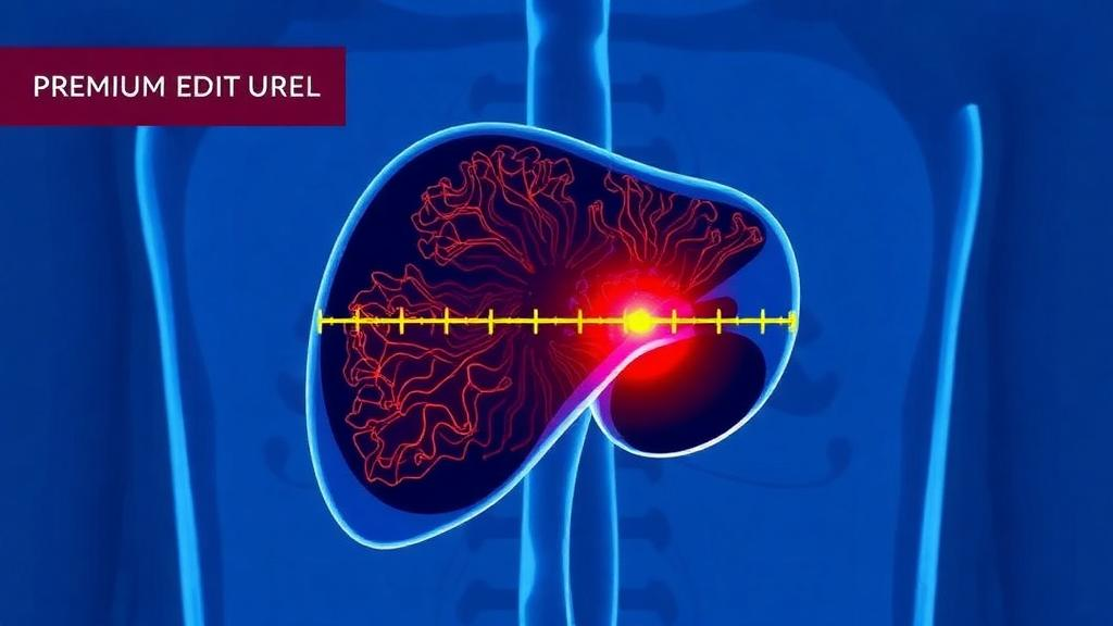
When doctors destroy a liver tumor with heat instead of surgery, the entire battle is won or lost by millimeters. Thermal ablation—using radiofrequency or microwave energy to cook a tumor in place—has become a mainstay of curative-intent treatment for small primary and secondary liver cancers, including hepatocellular carcinoma, intrahepatic cholangiocarcinoma, and colorectal liver metastases. Unlike surgical resection, where a pathologist can inspect the excised tissue under a microscope, ablation leaves the dead tumor in the body. The only way to judge whether the treatment killed every cancer cell is to compare images taken before and after the procedure and measure the rim of destroyed tissue, the so-called minimum ablative margin, that surrounds the tumor. A new simulation study published in CVIR Oncology now shows that the accuracy of that measurement itself may be the deciding factor in whether the widely used 5-millimeter margin standard can be trusted at all.
The research, led by Iwan Paolucci and colleagues at The University of Texas MD Anderson Cancer Center, tackled a deceptively simple question: how much do the errors built into ablation confirmation software distort the margins we think we are measuring? The concept of an A0 ablation—analogous to the R0 resection in surgery—demands complete tumor coverage with a pre-specified margin, conventionally at least 5 millimeters, and a correspondingly low risk of local tumor progression. But because the tumor is destroyed in situ, the true margin can never be measured histologically. Instead, clinicians rely on an imaging-based surrogate: they co-register pre-ablation and post-ablation scans, contour the tumor and the ablation zone, and compute the shortest three-dimensional distance between them. Every step of that pipeline introduces error, and until now there has been no systematic way to quantify how those errors propagate into the margin threshold that should actually be required.
The team built a mathematical and computational framework that models the entire measurement chain. They identified five key sources of inaccuracy from the literature—image resolution, segmentation error, registration error, tissue deformation, and image artifacts—and folded the last two into related parameters. Segmentation error, the mismatch between a drawn contour and the true boundary of the tumor or ablation zone, was modeled as random noise added to the contours, with tumor and ablation errors treated as independent. Registration error, the misalignment introduced when the two scans are aligned, was modeled as a shift of the ablation zone with a normally distributed magnitude and a random direction along the X, Y, or Z axis. Slice thickness, which ranges from 1 to 5 millimeters in published ablation confirmation studies, was simulated by resampling the synthetic images at different resolutions in the cranio-caudal direction, since in-plane resolution is typically already below 1 millimeter.
Crucially, the researchers also incorporated two biological effects that no imaging system can capture. The first is tissue shrinkage: microwave ablation causes radial contraction of tissue, with ex vivo experiments in bovine liver reporting contraction of up to 40 percent, an upper bound likely inflated by the absence of blood perfusion. Shrinkage within the ablation zone can make the measured margin appear larger than it truly is, and the exact contraction for any individual tumor cannot be observed during the procedure. The second effect is the presence of microscopic satellite lesions—tiny tumor deposits adjacent to the main lesion that fall below the roughly 1-millimeter spatial resolution of cross-sectional imaging. The model assumed these satellites are directly adjacent to the tumor, with sizes uniformly distributed between 0.5 and 2.5 millimeters, and with their presence governed by a binomial probability. Both effects bias the observed margin upward, meaning the measured number can flatter a treatment that has actually fallen short.
The scale of the simulation was enormous: 10,000 individual simulations for each of 1,500 parameter permutations, totaling 15 million synthetic ablation scenarios. In each run, the framework sampled a tumor size and margin, generated synthetic images of the tumor and ablation zone, applied tissue shrinkage and satellite lesions, measured the true margin, then applied registration error, segmentation noise, and slice-thickness resampling before measuring the observed margin. For each parameter combination, a logistic regression was fitted across observed margins ranging from minus 5 to plus 10 millimeters, and the A0 threshold was defined as the observed margin at which the probability of true complete microscopic tumor coverage reached at least 99 percent. The entire framework was implemented in Python and packaged into a freely available web application, allowing clinicians to enter the technical specifications of their own ablation confirmation software and obtain a software-specific A0 threshold.
The results carry a clear hierarchy of blame. Segmentation error emerged as the single most influential factor: with segmentation errors of 1, 3, and 5 millimeters, the required A0 thresholds rose to 3.4, 5.2, and 8.4 millimeters respectively. Registration error followed closely, with thresholds of 3.4, 4.9, and 7.0 millimeters for registration errors of 1, 3, and 5 millimeters. Slice thickness, by contrast, had a negligible effect, shifting the threshold by at most half a millimeter across the 1-to-5-millimeter range—a difference the authors attribute to simulation noise, since it falls below the resolution of the model itself. Among the biological effects, microscopic satellite lesions proved potent: the threshold climbed from 3.4 millimeters with no satellites to 5.8, 7.4, 7.9, and 7.7 millimeters as the probability of satellite presence rose from 25 to 100 percent. Tissue shrinkage worked in the opposite direction, lowering the required threshold from 3.4 millimeters with no shrinkage to 2.8, 2.2, and 1.8 millimeters at 10, 20, and 30 percent contraction.
The practical verdict concerns the sacred 5-millimeter rule. When both segmentation and registration errors were held at or below 3 millimeters, the simulated A0 threshold stayed at or below 5 millimeters, meaning the conventional criterion reliably guaranteed complete tumor coverage in at least 99 percent of cases. But once either error exceeded 3 millimeters, the required threshold climbed above 5 millimeters, and the standard criterion became unreliable—clinicians could believe they had achieved an adequate margin while microscopic disease survived. The study also exposed a subtle bias in the clinical literature: many retrospective studies exclude cases with visually judged registration errors above 3 millimeters before determining optimal margin thresholds. The simulations showed that this exclusion practice systematically lowers the apparent A0 threshold, and that the discrepancy grows as true registration error increases. The authors argue that studies must therefore disclose how many cases were excluded and why.
Why does this matter beyond the statistics? The minimum ablative margin has repeatedly been shown to be the most important predictor of local tumor progression after ablation, and a recent systematic review reinforced 5 millimeters as a minimum requirement while suggesting 10 millimeters as optimal. Yet the field suffers from high heterogeneity, likely driven by differences in measurement methodology and accuracy. Clinical studies capable of validating an A0 threshold for each software package are impractical: the packages evolve rapidly, and capturing the full variation in tumor sizes, margins, and errors would require sample sizes exceeding a thousand patients per comparison. Worse, the biological confounders are fundamentally unmeasurable in patients—microscopic satellites are known only probabilistically from histological studies, and tissue shrinkage only from ex vivo experiments. For rare tumor types particularly prone to satellites, such as intrahepatic cholangiocarcinoma, the necessary sample sizes are simply unattainable. In silico methods like this framework offer the only realistic route to technical validation before clinical deployment.
The authors are candid about the limitations of their approach. Tumors and ablation zones were modeled as spheres and ellipsoids, simplifications of irregular real-world anatomy. Interactions between error sources were ignored, even though a biomechanical deformable registration algorithm, for example, would likely perform worse when fed inaccurate segmentations, and intensity-based registration might suffer from the lower signal-to-noise ratio of thin-slice images. Errors were assumed to follow zero-mean normal distributions, implying no systematic bias—an assumption that may not hold for every commercial package. Registration types, whether rigid or deformable, were not distinguished because their behavior is heavily implementation-dependent. Even so, the study delivers a concrete benchmark: ablation confirmation software should achieve registration and segmentation errors of 3 millimeters or less before its 5-millimeter margin readout can be trusted. For a field increasingly reliant on artificial intelligence-driven contouring and automated margin assessment, that number is now the bar every developer, regulator, and interventional radiologist should be measuring against.
Subject of Research: Effects of measurement errors on minimum ablative margin thresholds in thermal ablation of liver tumors
Article Title: The effects of measurement errors on minimum ablative margins after thermal ablation of liver tumors: a simulation study
Article References: Paolucci, I., Albuquerque, J., Siddiqi, N. S., Jones, A. K., Brock, K. K., & Odisio, B. C. (2026). The effects of measurement errors on minimum ablative margins after thermal ablation of liver tumors: a simulation study. CVIR Oncology, 2(1), Article 1. https://doi.org/10.1007/s44343-025-00029-9
Image Credits: AI Generated
DOI: 10.1007/s44343-025-00029-9
Keywords: thermal ablation, liver tumors, minimum ablative margin, ablation confirmation software, segmentation error, registration error, simulation study, microwave ablation, colorectal liver metastases, hepatocellular carcinoma, image guidance, A0 margin
Cite Scienmag News
Nathaniel Bowman. (October 1, 2026). How Measurement Errors Could Undermine the 5-Millimeter Rule in Liver Cancer Ablation. Scienmag. https://scienmag.com/how-measurement-errors-could-undermine-the-5-millimeter-rule-in-liver-cancer-ablation/
Nathaniel Bowman. "How Measurement Errors Could Undermine the 5-Millimeter Rule in Liver Cancer Ablation." Scienmag, 1 October 2026, https://scienmag.com/how-measurement-errors-could-undermine-the-5-millimeter-rule-in-liver-cancer-ablation/. Accessed 4 October 2026.
Nathaniel Bowman. "How Measurement Errors Could Undermine the 5-Millimeter Rule in Liver Cancer Ablation." Scienmag. October 1, 2026. https://scienmag.com/how-measurement-errors-could-undermine-the-5-millimeter-rule-in-liver-cancer-ablation/


