


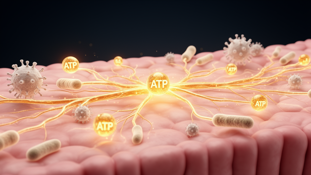
Deep inside the intestinal wall operates a nervous system so vast that scientists often call it the body’s second brain. The enteric nervous system contains between 200 and 600 million neurons, outnumbered several times over by star-shaped enteric glial cells, all woven together with the gut’s enormous immune apparatus and trillions of resident microbes. A new review published in Frontiers in Gastroenterology argues that the master switch coordinating this entire neuro-glial-immune axis may be something unexpectedly mundane: adenosine triphosphate, or ATP, the same molecule cells burn for energy. The work, led by researchers synthesizing decades of electrophysiology, pharmacology, and single-cell sequencing data, maps how purinergic signaling keeps the gut in balance and how its collapse may drive inflammatory bowel disease, dysmotility, and visceral pain.
The idea of purinergic transmission traces back to the 1970s, when Geoffrey Burnstock proposed that ATP itself acts as a signaling molecule released from nerves. The concept was initially met with skepticism, but it has since become a cornerstone of physiology. In the gut, ATP sits at unusually high intracellular concentrations of 1 to 10 millimoles per liter and escapes into the extracellular space through a menagerie of channels: vesicular exocytosis, volume-regulated anion channels, ATP-binding cassette transporters, connexins, and pannexins. Under healthy conditions, extracellular ATP hovers at nanomolar levels, quietly supporting mucosal barrier integrity and immune equilibrium. But infection, ischemia, and tissue injury unleash uncontrolled ATP floods that jump orders of magnitude, transforming the molecule into a danger-associated molecular pattern that recruits and activates T cells, macrophages, and mast cells.
Once outside the cell, ATP’s fate determines whether the signal calms or inflames. A family of ectonucleotidases sequentially dismantles it: NTPDases such as CD39 convert ATP to ADP and then AMP, and CD73 finishes the job by generating adenosine. This degradation cascade functions as a molecular switch between danger and resolution. ATP and its nucleotide derivatives activate P2 receptors, divided into ionotropic P2X channels and metabotropic P2Y receptors, while adenosine engages the four G protein-coupled P1 receptor subtypes A1, A2A, A2B, and A3. Each subtype carries distinct affinities, ranging from nanomolar for A1 and A2A to above one micromolar for A2B, and each couples to different intracellular cascades, granting the system remarkable combinatorial flexibility across cell types.
Within the enteric nervous system, purinergic transmission operates on three coordinated axes. ATP acts as a fast excitatory co-transmitter through P2X channels, generating rapid excitatory postsynaptic potentials that drive peristaltic reflexes. P2X3-expressing intrinsic primary afferent neurons, activated by ATP released from mucosal enterochromaffin cells, appear to sense luminal distension and launch the peristaltic reflex, and genetic deletion of P2X2 or P2X3 in mice measurably disrupts ileal peristalsis. Meanwhile, ATP acting at P2Y1 receptors on PDGFRalpha cells, which are electrically coupled to smooth muscle, generates inhibitory junction potentials that relax the gut wall. Adenosine provides a third, tonic braking influence, inhibiting acetylcholine release and motility through A1 and A2A receptors. Human tissue studies add a distinctive wrinkle: P2X1 is highly expressed and frequently co-localized with P2X2 in human submucosal neurons, a pattern not previously reported in other species.
Perhaps the most striking discovery is how intimately glia participate. Stimulating a single enteric neuron releases ATP through pannexin channels that triggers calcium responses in just two or three adjacent glial cells, revealing that neuron-glia communication is strikingly localized. Activated glia can then feed the signal forward, releasing more ATP through connexin-43 hemichannels and amplifying the original message. During intestinal inflammation, this loop turns destructive. Neuronal P2X7 receptors stimulate glial P2Y1 receptors, activated glia produce nitric oxide and release more ATP through connexin-43, and the accumulating extracellular ATP reactivates neuronal P2X7, creating a self-perpetuating cycle that ultimately kills enteric neurons, a mechanism the review links to inflammation-associated motility disorders.
On the immune side, no receptor has attracted more attention than P2X7. Expressed on macrophages, dendritic cells, T cells, neurons, and glia, P2X7 activates NF-kappaB, NFAT, and HIF1alpha transcriptional programs, upregulates TNF-alpha, IL-6, CCL2, and CXCL2, and stands among the most potent activators of the NLRP3 inflammasome, driving IL-1beta release. P2X7 expression is elevated in the epithelium and lamina propria of both murine colitis models and human IBD biopsies. In mice, genetic deletion or pharmacological blockade of P2X7 protects against chemically induced colitis, reducing immune cell infiltration and pro-inflammatory cytokines. Yet the review’s authors repeatedly caution that receptor abundance in human tissue demonstrates association, not causation, and whether these preclinical benefits translate to patients remains an open question.
Adenosine receptors generally play the opposite hand, though with crucial exceptions. A2A activation in T cells suppresses pro-inflammatory cytokine release and promotes regulatory T cell expansion, and A2A agonists reduce inflammation in murine colitis models. A3 agonists suppress NF-kappaB signaling in human intestinal epithelial cells and improve experimental colitis outcomes. But the A2B receptor can be frankly pro-inflammatory in chronic inflammation, and its effects on glia differ from those on epithelium. Even the ectonucleotidases resist simple classification: CD73 deletion worsens colitis in one mouse model, while pharmacological CD73 blockade alleviates it in another, and P2X4 signaling has been reported as both protective and pathogenic depending on the model, reflecting the system’s profound dependence on cell type, timing, and context.
The therapeutic implications are considerable but double-edged. Clinical correlates already exist: plasma ADPase activity, reflecting NTPDase-2 and NTPDase-3 function, is significantly reduced in Crohn’s disease patients and correlates with disease activity, while higher NTPDase-1 expression on regulatory T cells tracks with remission in IBD. P2Y14 gene expression in ulcerative colitis biopsies rises with disease severity, and P2Y14 antagonism attenuates experimental colitis in mice. But because purinergic signaling simultaneously governs motility, secretion, barrier maintenance, and host defense, systemically drugging a single receptor risks collateral damage to essential intestinal functions. The authors argue that future strategies must prioritize receptor selectivity and targeted delivery to specific cell populations.
What emerges is a picture of the gut as an organ run by an information economy in which ATP is simultaneously currency and alarm signal, its concentration and metabolism deciding whether neurons fire, glia relay, epithelia secrete, or immune cells attack. The review calls for single-cell and spatial transcriptomics, functional imaging, and rigorous validation in human intestinal tissues to separate genuine disease drivers from adaptive responses to inflammation. If that effort succeeds, the purinergic system could yield new biomarkers and treatments for inflammatory bowel disease, post-inflammatory dysmotility, and visceral pain syndromes, ailments that currently afflict millions worldwide with limited therapeutic options.
Subject of Research: Purinergic signaling in the gut neuro-glial-immune axis and its role in intestinal homeostasis and inflammatory bowel disease
Article Title: Purinergic signaling in the gut neuro-glial-immune axis: mechanisms of homeostasis, inflammation, and gastrointestinal dysfunction
Article References: Valdez-Morales, E. E., Liñán-Rico, A., & Guerrero-Alba, R. (2026). Purinergic signaling in the gut neuro-glial-immune axis: mechanisms of homeostasis, inflammation, and gastrointestinal dysfunction. Frontiers in Gastroenterology, 5, Article 1954794. https://doi.org/10.3389/fgstr.2026.1954794
Image Credits: AI Generated
DOI: 10.3389/fgstr.2026.1954794
Keywords: purinergic signaling, ATP, enteric nervous system, enteric glial cells, inflammatory bowel disease, P2X7 receptor, adenosine receptors, ectonucleotidases, gut microbiota, neuroimmune communication, intestinal motility, visceral pain
Cite Scienmag News
Morgan Morrow. (October 16, 2026). How ATP Wires the Gut’s Brain, Immune System and Microbiome Into One Circuit. Scienmag. https://scienmag.com/how-atp-wires-the-guts-brain-immune-system-and-microbiome-into-one-circuit/
Morgan Morrow. "How ATP Wires the Gut’s Brain, Immune System and Microbiome Into One Circuit." Scienmag, 16 October 2026, https://scienmag.com/how-atp-wires-the-guts-brain-immune-system-and-microbiome-into-one-circuit/. Accessed 16 October 2026.
Morgan Morrow. "How ATP Wires the Gut’s Brain, Immune System and Microbiome Into One Circuit." Scienmag. October 16, 2026. https://scienmag.com/how-atp-wires-the-guts-brain-immune-system-and-microbiome-into-one-circuit/


